 Français
 English
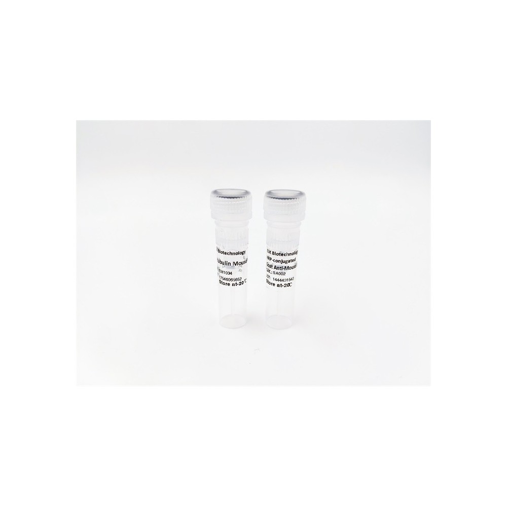

| ELK.No | ES8510 |
| Product name | Perforin 1 rabbit pAb |
| Reactivity | Human;Rat;Mouse; |
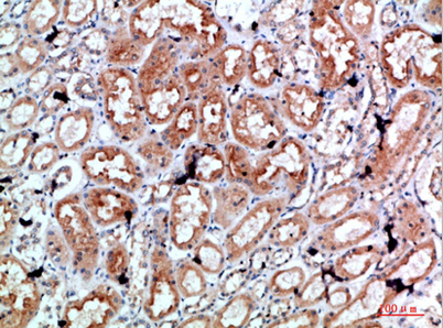
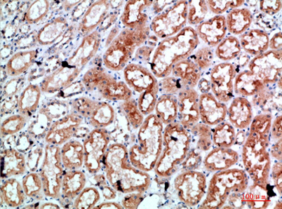
| Applications | WB;IHC;ELISA |
| Other name | Perforin-1 (P1) (Cytolysin) (Lymphocyte pore-forming protein) (PFP) |
| Size | 100μL |
| Unit price ($) | 248 |
| Human gene ID | 5551 |
| Human Swiss-Prot | P14222 |
| Source | Rabbit |
| Isotype | IgG |
| Target | Perforin |
| Fields | >>Apoptosis;>>Natural killer cell mediated cytotoxicity;>>Type I diabetes mellitus;>>Autoimmune thyroid disease;>>Allograft rejection;>>Graft-versus-host disease;>>Viral myocarditis |
| Gene name | PRF1 |
| Protein name | Perforin 1 |
| Human gene link | View Human Gene |
| Human Swiss link | View Human Swiss-Prot |
| Mouse gene ID | 18646 |
| Mouse gene link | |
| Mouse Swiss-Prot | P10820 |
| Mouse Swiss link | View Mouse Swiss-Prot |
| Rat gene ID | |
| Rat gene link | |
| Rat Swiss-Prot | |
| Rat Swiss link | |
| Immunogen | The antiserum was produced against synthesized peptide derived from the C-terminal region of human PRF1. AA range:451-500 |
| Specificity | Perforin 1 Polyclonal Antibody detects endogenous levels of Perforin 1 |
| Formulation | Liquid in PBS containing 50% glycerol, 0.5% BSA and 0.02% sodium azide. |
| Clonality | Polyclonal |
| Dilution | WB 1:500-2000, IHC 1:50-200, ELISA 1:10000-20000 |
| Purification | The antibody was affinity-purified from rabbit antiserum by affinity-chromatography using epitope-specific immunogen. |
| Concentration | 1 mg/ml |
| Storage stability | -20°C/1 year |
| Molecular Weight (Da) | |
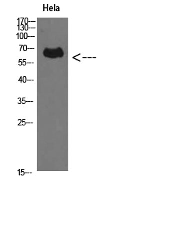
| Observed band (KD) | 61kD |
| Background | The protein encoded by this gene has structural and functional similarities to complement component 9 (C9). Like C9, this protein creates transmembrane tubules and is capable of lysing non-specifically a variety of target cells. This protein is one of the main cytolytic proteins of cytolytic granules, and it is known to be a key effector molecule for T-cell- and natural killer-cell-mediated cytolysis. Defects in this gene cause familial hemophagocytic lymphohistiocytosis type 2 (HPLH2), a rare and lethal autosomal recessive disorder of early childhood. Alternative splicing results in multiple transcript variants encoding the same protein. [provided by RefSeq, Jul 2008], |
| Function | disease:Defects in PRF1 are the cause of familial hemophagocytic lymphohistiocytosis type 2 (FHL2) [MIM:603553]; also known as HPLH2. Familial hemophagocytic lymphohistiocytosis (FHL) is a genetically heterogeneous, rare autosomal recessive disorder. It is characterized by immune dysregulation with hypercytokinemia and defective natural killer cell function. The clinical features of the disease include fever, hepatosplenomegaly, cytopenia, hypertriglyceridemia, hypofibrinogenemia, and neurological abnormalities ranging from irritability and hypotonia to seizures, cranial nerve deficits, and ataxia. Hemophagocytosis is a prominent feature of the disease, and a non-malignant infiltration of macrophages and activated T lymphocytes in lymph nodes, spleen, and other organs is also found.,function:In the presence of calcium, perforin polymerizes into transmembrane tubules and is capable of lys |
| Subcellular location | Cytolytic granule . Secreted. Cell membrane ; Multi-pass membrane protein . Endosome lumen . Stored in cytolytic granules of cytolytic T-lymphocytes and secreted into the cleft between T-lymphocyte and target cell (PubMed:20038786). Inserts into the cell membrane of target cells and forms pores (PubMed:20889983). Membrane insertion and pore formation requires a major conformation change (PubMed:20889983). May be taken up via endocytosis involving clathrin-coated vesicles and accumulate in a first time in large early endosomes (PubMed:20038786). . |
| Expression | Liver,Natural killer cell,Spleen, |

Use collapsible tabs for more detailed information that will help customers make a purchasing decision.
Ex: Shipping and return policies, size guides, and other common questions.