 Français
 English
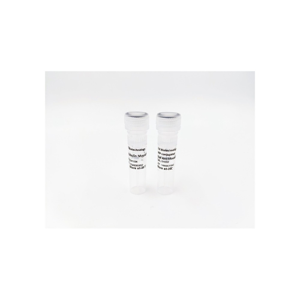

| ELK.No | ES8791 |
| Product name | CD274 rabbit pAb |
| Reactivity | Human;Rat;Mouse; |
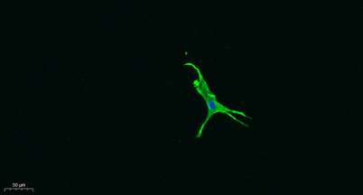
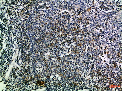
| Applications | IHC;IF;ELISA |
| Other name | Programmed cell death 1 ligand 1 (PD-L1) (PDCD1 ligand 1) (Programmed death ligand 1) (B7 homolog 1) (B7-H1) (CD antigen CD274) |
| Size | 50μL |
| Unit price ($) | 148 |
| Human gene ID | 29126 |
| Human Swiss-Prot | Q9NZQ7 |
| Source | Rabbit |
| Isotype | IgG |
| Target | CD274 |
| Fields | >>Cell adhesion molecules;>>PD-L1 expression and PD-1 checkpoint pathway in cancer |
| Gene name | CD274 B7H1 PDCD1L1 PDCD1LG1 PDL1 |
| Protein name | Programmed cell death 1 ligand 1 (PD-L1) (PDCD1 ligand 1) (Programmed death ligand 1) (B7 homolog 1) (B7-H1) (CD antigen CD274) |
| Human gene link | View Human Gene |
| Human Swiss link | View Human Swiss-Prot |
| Mouse gene ID | 60533 |
| Mouse gene link | |
| Mouse Swiss-Prot | Q9EP73 |
| Mouse Swiss link | View Mouse Swiss-Prot |
| Rat gene ID | |
| Rat gene link | |
| Rat Swiss-Prot | |
| Rat Swiss link | |
| Immunogen | Synthetic peptide from human protein at AA range: 181-230 |
| Specificity | The antibody detects endogenous CD274 |
| Formulation | Liquid in PBS containing 50% glycerol, 0.5% BSA and 0.02% sodium azide. |
| Clonality | Polyclonal |
| Dilution | IHC-p 1:50-200, ELISA 1:10000-20000 IF 1:100-300 Not yet tested in other applications. |
| Purification | The antibody was affinity-purified from rabbit antiserum by affinity-chromatography using epitope-specific immunogen. |
| Concentration | 1 mg/ml |
| Storage stability | -20°C/1 year |
| Molecular Weight (Da) | |
| Observed band (KD) | |
| Background | This gene encodes an immune inhibitory receptor ligand that is expressed by hematopoietic and non-hematopoietic cells, such as T cells and B cells and various types of tumor cells. The encoded protein is a type I transmembrane protein that has immunoglobulin V-like and C-like domains. Interaction of this ligand with its receptor inhibits T-cell activation and cytokine production. During infection or inflammation of normal tissue, this interaction is important for preventing autoimmunity by maintaining homeostasis of the immune response. In tumor microenvironments, this interaction provides an immune escape for tumor cells through cytotoxic T-cell inactivation. Expression of this gene in tumor cells is considered to be prognostic in many types of human malignancies, including colon cancer and renal cell carcinoma. Alternative splicing results in multiple transcript variants. [provided by RefSeq, Sep 2015], |
| Function | function:Involved in the costimulatory signal, essential for T-cell proliferation and production of IL10 and IFNG, in an IL2-dependent and a PDCD1-independent manner. Interaction with PDCD1 inhibits T-cell proliferation and cytokine production.,induction:Up-regulated on T- and B-cells, dendritic cells, keratinocytes and monocytes after LPS and IFNG activation. Up-regulated in B-cells activated by surface Ig cross-linking.,similarity:Belongs to the immunoglobulin superfamily. BTN/MOG family.,similarity:Contains 1 Ig-like C2-type (immunoglobulin-like) domain.,similarity:Contains 1 Ig-like V-type (immunoglobulin-like) domain.,subunit:Interacts with PDCD1.,tissue specificity:Highly expressed in the heart, skeletal muscle, placenta and lung. Weakly expressed in the thymus, spleen, kidney and liver. Expressed on activated T- and B-cells, dendritic cells, keratinocytes and monocytes., |
| Subcellular location | Cell membrane ; Single-pass type I membrane protein . Early endosome membrane ; Single-pass type I membrane protein . Recycling endosome membrane ; Single-pass type I membrane protein . Associates with CMTM6 at recycling endosomes, where it is protected from being targeted for lysosomal degradation. .; [Isoform 1]: Cell membrane ; Single-pass type I membrane protein .; [Isoform 2]: Endomembrane system ; Single-pass type I membrane protein . |
| Expression | Highly expressed in the heart, skeletal muscle, placenta and lung. Weakly expressed in the thymus, spleen, kidney and liver. Expressed on activated T- and B-cells, dendritic cells, keratinocytes and monocytes. |

Use collapsible tabs for more detailed information that will help customers make a purchasing decision.
Ex: Shipping and return policies, size guides, and other common questions.