 Français
 English
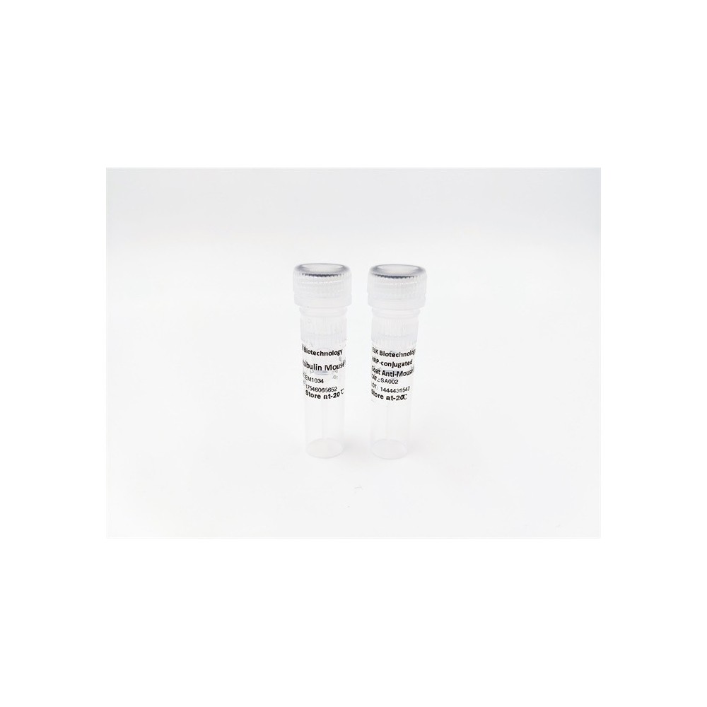

| ELK.No | ES9692 |
| Product name | REM1 rabbit pAb |
| Reactivity | Human;Mouse |
| Applications | WB;ELISA |
| Other name | |
| Size | 50μL |
| Unit price ($) | 148 |
| Human gene ID | 28954 |
| Human Swiss-Prot | O75628 |
| Source | Rabbit |
| Isotype | IgG |
| Target | REM1 |
| Fields | |
| Gene name | REM1 GES REM |
| Protein name | GTP-binding protein REM 1 (GTPase-regulating endothelial cell sprouting) (Rad and Gem-like GTP-binding protein 1) |
| Human gene link | |
| Human Swiss link | View Human Swiss-Prot |
| Mouse gene ID | |
| Mouse gene link | |
| Mouse Swiss-Prot | O35929 |
| Mouse Swiss link | View Mouse Swiss-Prot |
| Rat gene ID | |
| Rat gene link | |
| Rat Swiss-Prot | |
| Rat Swiss link | |
| Immunogen | Synthesized peptide derived from part region of human protein |
| Specificity | REM1 Polyclonal Antibody detects endogenous levels of protein. |
| Formulation | Liquid in PBS containing 50% glycerol, and 0.02% sodium azide. |
| Clonality | Polyclonal |
| Dilution | WB 1:500-2000 ELISA 1:5000-20000 |
| Purification | The antibody was affinity-purified from rabbit antiserum by affinity-chromatography using epitope-specific immunogen. |
| Concentration | 1 mg/ml |
| Storage stability | -20°C/1 year |
| Molecular Weight (Da) | |
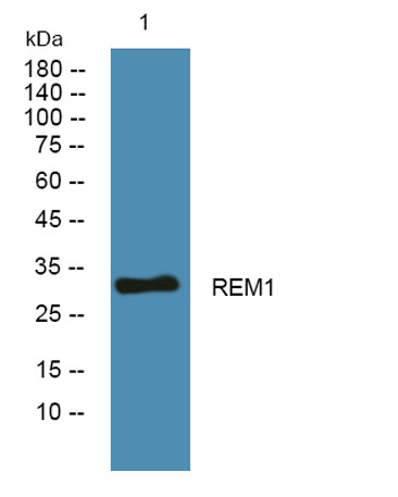
| Observed band (KD) | 32kD |
| Background | The protein encoded by this gene is a GTPase and member of the RAS-like GTP-binding protein family. The encoded protein is expressed in endothelial cells, where it promotes reorganization of the actin cytoskeleton and morphological changes in the cells. [provided by RefSeq, Jul 2008], |
| Function | function:Promotes endothelial cell sprouting and actin cytoskeletal reorganization. May be involved in angiogenesis. May function in Ca(2+) signaling.,similarity:Belongs to the small GTPase superfamily. RGK family.,subunit:In vitro, interacts with calmodulin in a calcium-dependent manner.,tissue specificity:Most highly expressed in the endothelial lining of the blood vessels in uterus and heart. Lower levels found in spleen, lymph node, kidney and testis. Also found in cells with secretory function such as the islets of Langerhans, lobule/duct epithelium in the breast, bile duct epithelium in the liver, surface epithelium in the endometrial glands of the uterus, colon mucosa and acinar cells in the pancreas and the prostate., |
| Subcellular location | intracellular,membrane, |
| Expression | Most highly expressed in the endothelial lining of the blood vessels in uterus and heart. Lower levels found in spleen, lymph node, kidney and testis. Also found in cells with secretory function such as the islets of Langerhans, lobule/duct epithelium in the breast, bile duct epithelium in the liver, surface epithelium in the endometrial glands of the uterus, colon mucosa and acinar cells in the pancreas and the prostate. |

Use collapsible tabs for more detailed information that will help customers make a purchasing decision.
Ex: Shipping and return policies, size guides, and other common questions.
