 Français
 English

| ELK.No | ES9851 |
| Product name | MYO1F rabbit pAb |
| Reactivity | Human;Mouse |
| Applications | WB;ELISA |
| Other name | |
| Size | 50μL |
| Unit price ($) | 148 |
| Human gene ID | 4542 |
| Human Swiss-Prot | O00160 |
| Source | Rabbit |
| Isotype | IgG |
| Target | MYO1F |
| Fields | >>Pathogenic Escherichia coli infection |
| Gene name | MYO1F |
| Protein name | Unconventional myosin-If (Myosin-Ie) |
| Human gene link | |
| Human Swiss link | View Human Swiss-Prot |
| Mouse gene ID | |
| Mouse gene link | |
| Mouse Swiss-Prot | P70248 |
| Mouse Swiss link | View Mouse Swiss-Prot |
| Rat gene ID | |
| Rat gene link | |
| Rat Swiss-Prot | |
| Rat Swiss link | |
| Immunogen | Synthesized peptide derived from human protein . at AA range: 640-720 |
| Specificity | MYO1F Polyclonal Antibody detects endogenous levels of protein. |
| Formulation | Liquid in PBS containing 50% glycerol, and 0.02% sodium azide. |
| Clonality | Polyclonal |
| Dilution | WB 1:500-2000 ELISA 1:5000-20000 |
| Purification | The antibody was affinity-purified from rabbit antiserum by affinity-chromatography using epitope-specific immunogen. |
| Concentration | 1 mg/ml |
| Storage stability | -20°C/1 year |
| Molecular Weight (Da) | |
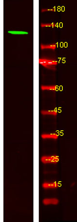
| Observed band (KD) | 120kD |
| Background | function:Myosins are actin-based motor molecules with ATPase activity. Unconventional myosins serve in intracellular movements. Their highly divergent tails are presumed to bind to membranous compartments, which would be moved relative to actin filaments.,similarity:Contains 1 IQ domain.,similarity:Contains 1 myosin head-like domain.,similarity:Contains 1 SH3 domain., |
| Function | function:Myosins are actin-based motor molecules with ATPase activity. Unconventional myosins serve in intracellular movements. Their highly divergent tails are presumed to bind to membranous compartments, which would be moved relative to actin filaments.,similarity:Contains 1 IQ domain.,similarity:Contains 1 myosin head-like domain.,similarity:Contains 1 SH3 domain., |
| Subcellular location | myosin complex,unconventional myosin complex, |
| Expression | Blood,Brain,Epithelium,Retina,Spleen, |

Use collapsible tabs for more detailed information that will help customers make a purchasing decision.
Ex: Shipping and return policies, size guides, and other common questions.
