Anti-Cathepsin V Rabbit pAb - 50 µL

AO-03-GB112712-50
  • Specification: 50 µL
  • Applications: IHC | IF
  • Species: H,M,R | H,M,R
  • Dilution: 1: 1000-1: 2000 | 1: 300-1: 600
  • Positive tissue: kidney, duodenum | kidney
check In Stock
Hurry! only 10 items left in stock.
€299.00
Tax excluded
Quantity

Product Information

Protein full name

Cathepsin L2

Synonyms

cathepsin L2, Cathepsin U, Cathepsin V, CATL2, CTSL2, CTSU, CTSV

Uniprot ID

O60911

Immunogen

Recombinant protein corresponding to Human Cathepsin V

Isotype

IgG

Purity

Affinity purification

Subcellular location

Lysosome

Applications

ApplicationsSpeciesDilutionPositive Tissue
IHC/IFHuman, Mouse, Rat1: 300-1: 2000kidney

Background

Cathepsin V is a lysosomal cysteine protease which has an important role in corneal physiology.Cathepsin V was found to be differentially expressed in relation to pigmentary skin backgrounds, relative to darkly pigmented skins. It is expressed in primary keratinocytes, melanocytes, and HaCaT keratinocytes, but not in primary fibroblasts. The purified cathepsin L2 was a high molecular weight proteinase of about 78 kDa, but it was unstable to SDS and reducing conditions, which dissociated parts of the proteinase into 66 kDa, 31 kDa and 26 kDa fragments and it can form a complex of 70 kDa with an unidentified subunit.

Images

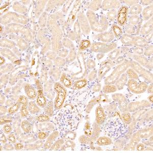
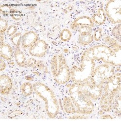
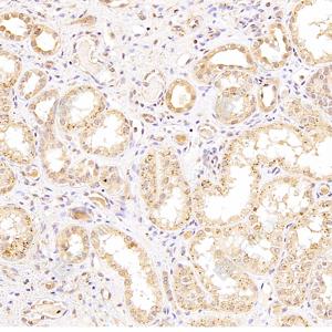
01ywmnQsLKqT8pJ.jpg

IHC analysis of Cathepsin V(GB112712).
Sample: Human kidney, 4% PFA (G1101) 12-24h.
Antigen retrieval: Citrate buffer (pH 6.0) (G1201), 98°C, 20 min.
Primary antibody: 1: 300, 4°C overnight.
Secondary antibody: HRP conjugated Goat Anti-Rabbit lgG (H+L) (GB23303), 1: 200, RT, 1h.

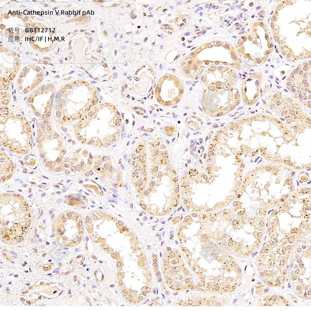
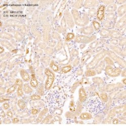
02U4EGztPmbenw9.jpg

IHC analysis of Cathepsin V(GB112712).
Sample: Mouse kidney, 4% PFA (G1101) 12-24h.
Antigen retrieval: Citrate buffer (pH 6.0) (G1201), 98°C, 20 min.
Primary antibody: 1: 700, 4°C overnight.
Secondary antibody: HRP conjugated Goat Anti-Rabbit lgG (H+L) (GB23303), 1: 200, RT, 1h.

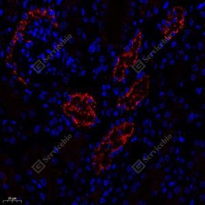
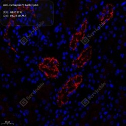
03f9Vqx2wsEEypr.jpg

IF analysis of Cathepsin V(GB112712)red.
Sample: Mouse kidney, 4% PFA (G1101) 12-24h.
Antigen retrieval: Tris-EDTA buffer (pH 9.0) (G1203), 98°C, 20min.
Blocking buffer: 3% BSA (GC305010) in PBS, RT, 30min.
Primary antibody: 1: 600, 4°C overnight.
Secondary antibody: Cy3 conjugated Goat Anti-Rabbit IgG (H+L) (GB21303), 1: 300, RT, 1h.

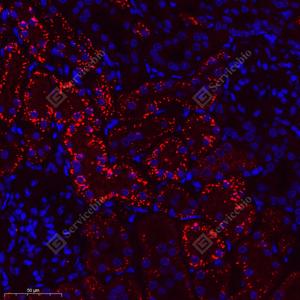
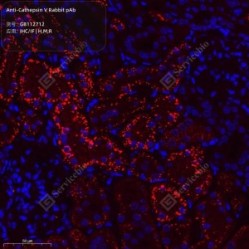
04KPFQY7h47KjHp.jpg

IF analysis of Cathepsin V(GB112712)red.
Sample: Rat kidney, 4% PFA (G1101) 12-24h.
Antigen retrieval: Tris-EDTA buffer (pH 9.0) (G1203), 98°C, 20min.
Blocking buffer: 3% BSA (GC305010) in PBS, RT, 30min.
Primary antibody: 1: 600, 4°C overnight.
Secondary antibody: Cy3 conjugated Goat Anti-Rabbit IgG (H+L) (GB21303), 1: 300, RT, 1h.

Storage

Storage Store at -20°C for one year. Avoid repeated freeze/thaw cycles.
Storage Buffer PBS with 0.15% ProClin300, 100 µg/mL BSA and 50% glycerol.

NOTE:

1.This product is intended for research only.

2.This product is recommended to dilute with the Primary Antibody Dilution Buffer (G2025).

Datasheet: Download datasheet 1

: AO-03-GB112712-50
: 10 Items
Hurry! only 10 items left in stock.

Use collapsible tabs for more detailed information that will help customers make a purchasing decision.

Ex: Shipping and return policies, size guides, and other common questions.

  • Paste the label on a flat surface on the package
  • Make sure that both 1D and 2D barcodes are clearly visible
  • Ensure that the label is smooth and isn’t creased or wrinkled
  • Check for any tears, dents, holes or scratches
  • Pack your product tightly, with the right size packaging
  • Ensure both barcodes are on a flat surface of the package