Product Information
|
Product Name
|
Cat.No.
|
Spec.
|
|
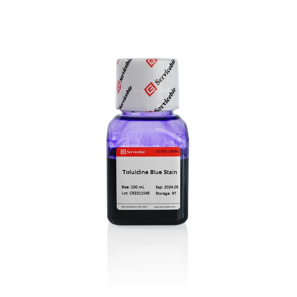
Toluidine Blue Dye Solution
|
AO-03-G1032-100ML
|
100 mL
|
Description
Toluidine Blue is a quinone imine basic staining, which can combine with acidic substances in tissue cells to achieve tissue staining. The nuclei and Nissl's body in neurons is stained into blue with toluidine blue, which can be used for the preliminary pathological diagnosis of Nissl body; The cytoplasm of mast cells contains heparin and histamine, and the cartilage contains chondroitin sulfate. These substances have metachromatic properties and are stained intopurplish red with toluidine blue. Therefore, toluidine blue staining can be used to observe the distribution and abnormal changes of mast cells and the morphological structure of cartilage, such as tide lines; plant tissues can be stained and observed, the xylem, ducts, sieve tubes and other structures, the xylem arestained into blue-green and the cellulose cell wallis stained into blue-purple.
The active ingredient concentration of the toluidine blue dye solution of this product is 0.5%, which can be used for the staining of conventional animal and plant tissue sections.
Storage and Handling Conditions
Transport at room temperature; Valid for 18 months.
Component
|
Component
|
AO-03-G1032
|
|
Toluidine Blue Dye Solution
|
100 mL
|
|
Manual
|
1 pc
|
Assay Protocol / Procedures
1. Paraffin sections were dewaxed to water;
2. Toluidine blue stain:Put the tissue sections into the toluidine blue dye solution for 2-5 minutes, wash slightly with tap water to remove excess dye.
a. For plant tissue: Controll the degree of staining under the microscope,and according to the degree of staining, use 0.1% glacial acetic acid for proper differentiation. If the degree of coloring is appropriate and does not require differentiation, the slices are dried in a 60°C oven.
b. For animal tissue: Wash animal tissue sections with water and differentiated with 0.1% glacial acetic acid. The degree of differentiation was observed microscopically, and differentiation was sufficient until the background was light blue and the coloration of the nidus, cartilage, and mast cells was evident. Rinse with tap water to terminate differentiation and then dry it in an oven at 60℃.
3. Transparent and mount:Sections were transparent to xylene for 10 min and then sealed with neutral gum.
Note
1. The washing time of the stained sections should not be too long, otherwise the color will fade easily.
2. Sections must be completely dried prior to transparent sealing so that any remaining tiny droplets of water do not interfere with observation.
3. Staining Solution is reusable. 100 mL of Staining Solution can be used to stain (dip or drop) approximately 350 sections. Replace the stain with a new one when the tissue or cell coloring is significantly lighter or abnormal.
4. Please wear lab coat and disposable gloves during operation.
For Research Use Only!