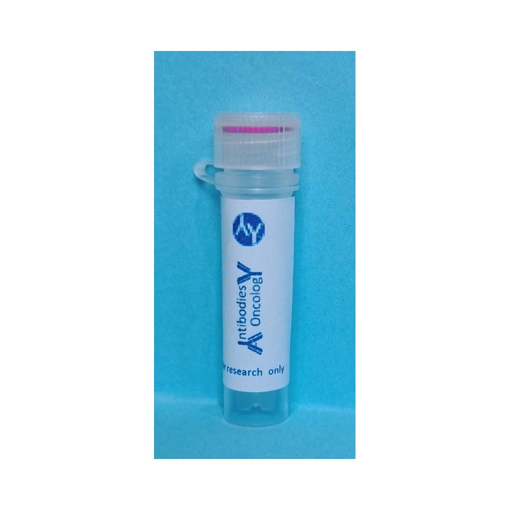


Product Information
|
Product Name |
Cat.No. |
Spec.. |
|
Calcein AM |
AO-03-G1728 |
0.1 mL |
Product Description/Introduction
Calcein AM (Calcein acetoxymethyl ester), is based on Calcein with the introduction of acetoxymethyl ester (AM) group, which increases the hydrophobicity, so it can easily penetrate the membrane of the living cells. Calcein AM itself is a cell-permeable, non-fluorescent and hydrophobic compound that, upon entry into the cell, is hydrolyzed by endogenous esterases in the cell to produce the strongly negatively charged polar molecule Calcein that cannot permeate the cell membrane, and thus is retained within the cell, whereas Calcein (maximum excitation wavelength: 494 nm; maximum emission wavelength: 517 nm) can emit strong green fluorescence. Compared to other similar probes (e.g. BCECF AM and CFDA AM), Calcein AM is currently one of the most ideal fluorescent probes for staining live cells due to its very low cytotoxicity, virtually no effect on cellular functions such as cell proliferation or lymphocyte chemotaxis, and low pH sensitivity.
Due to the lack of esterase in dead cells, Calcein AM is only used for viability testing and short-term labeling of living cells. The red fluorescent nucleic acid dye Propidium Iodide (PI) does not penetrate the cell membrane of living cells, it passes through the disordered region of the dead cell membrane to the nucleus and embeds itself in the cell's DNA double helix to produce a red fluorescence (excitation: 535 nm, emission: 617 nm), therefore PI stains only dead cells. Calcein AM is often used in combination with propidium iodide (PI)for simultaneous dual fluorescence staining of live and dead cells. Since both Calcein and PI-DNA can be excited at 490 nm, live and dead cells can be observed simultaneously by fluorescence microscopy. With 545 nm excitation, only dead cells can be observed.
Calcein AM can be used in most mammalian cells. It has been shown that Calcein AM can be used in some plant cells such as Arabidopsis root margin-like cells and some yeasts such as Pichia anomala and Saccharomyces cerevisiae. Certain parasites such as Leishmania cannot enter living cells due to cell membrane components, but Calcein AM can enter parasite cells in the early stages of apoptosis, and thus is used in conjunction with PI for the detection of parasites in the early stages of apoptosis. Calcein AM is not suitable for use on fungi and bacteria because they have cell walls that prevent Calcein AM from entering cells.
Calcein itself is a metal complexation indicator, and the fluorescent signal is quenched when it complexes with metal ions such as Co2+, Ni2+, Cu2+, Fe3+, and Mn2+ at physiological pH, making Calcein AM also a green fluorescent probe that can be used to determine the mitochondrial permeability transition pore.
This product, Calcein AM, is of high purity and dissolved in anhydrous DMSO at a concentration of 2 mM.The commonly used final concentration of Calcein AM is 0.1-5.0 μM.In order to obtain a more desirable result, please adjust the final concentration of Calcein appropriately according to the type of cells and the actual situation of the experiment.
Storage and Shipping Conditions
-20℃ for storage and transportation, valid for 12 months.
ProductContent
|
Component |
AO-03-G1728 |
|
Calcein AM |
0.1 mL |
|
Manual |
1 pc |
Assay Protocol / Procedures
Calcein AM staining working solution was prepared:
Dilute the product with a suitable buffer, such as serum-free culture medium, HBSS (G4203), or PBS (G4202), to prepare a Calcein AM staining working solution at a concentration of 0.1-5.0 µM.
Note: The final concentration of Calcein AM is recommended to be adjusted appropriately according to the cell type and experimental reality.
Procedures
1. Fluorescence microscopy assay or fluorescence zymography assay for suspension cells:
a) Cells were counted after certain treatments according to the experimental design. Take the appropriate cells and centrifuge them at 250×g for 5 min at room temperature, discard the supernatant, and add the appropriate volume of Calcein AM staining working solution to make the cell density about 1×10⁶/ml.
b) Incubate the cells at 37ºC for 30-45 min, the optimal incubation time is different for different cells. Consider 30 min as the initial incubation time, and optimize it according to the specific experimental situation to get more ideal detection results.
c) At the end of incubation, centrifuge at 250×g for 5 min, aspirate the supernatant, and slowly resuspend the cells by adding 37ºC pre-warmed culture medium.
d) Repeat step c two or more times to adequately wash to remove residual staining solution.
e) Replace the fresh 37ºC pre-warmed culture medium and incubate at 37ºC for another 30 minutes away from light to ensure that the intracellular esterases sufficiently hydrolyze Calcein AM to produce Calcein with green fluorescence.
f) The cells were centrifuged at 250×g for 5 min at room temperature, most of the culture medium was aspirated, and the cells were resuspended with the remaining culture medium and smeared, and then observed under a fluorescence microscope or detected by a fluorescence zymography.The maximal excitation wavelength of Calcein is 494 nm, and the maximal emission wavelength is 514 nm.If necessary, other fluorescence re-staining can be carried out, and the whole process should be avoided by light operation.
2. Fluorescence microscopy assay of adherent cells or fluorescence enzyme labeling assay:
a) Cells were inoculated on petri dishes, multiwell cell culture plates or cell crawlers and treated in certain ways according to the experimental design.
b) Aspirate the culture solution and wash the cells 1-2 times with PBS.
c) Add an appropriate volume of Calcein AM Staining Solution and shake gently so that the dye covers all cells evenly. Generally, the volume of 96-well plate is 100 μl per well, 24-well plate is 250 μl per well, 12-well plate is 500 μl per well, and 6-well plate is 1 ml per well.
d) Incubate at 37ºC for 30-45 min, the optimal incubation time varies for different cells. Take 30 min as the initial incubation time, and optimize the incubation time according to the cells used to get more ideal staining effect.
e) At the end of the incubation, the culture medium was replaced with fresh pre-warmed culture medium at 37ºC and incubated at 37ºC for another 30 min away from light to ensure that the intracellular esterase sufficiently hydrolyzed Calcein AM to produce Calcein with green fluorescence.
f) Aspirate the culture fluid, wash with PBS for 2-3 times, and then add the serum-free cell culture fluid can be observed under a fluorescence microscope or detected by fluorescent enzyme marker.Calcein has a maximum excitation wavelength of 494 nm and a maximum emission wavelength of 514 nm, and can be further stained with other fluorescents if desired, keeping the entire process away from light.
3. Flow cytometry assay:
a) After trypsin digestion of adherent cells, resuspend the cells in culture medium, suspend the cells for direct use, count, centrifuge an appropriate amount of cells at 250×g for 5 min at room temperature, discard the supernatant, and add an appropriate volume of Calcein AM staining working solution so that the cells are a single-cell suspension and the density of the cells is approximately 1×10⁶ /ml, with a volume of 1 ml for each sample. NOTE: It is necessary to prepare a buffer-only sample of the cells for use as a negative control in the flow cytometry assay. negative control for the flow cytometry assay, and it is desirable to keep this buffer the same as the buffer used to prepare the Calcein AM Staining Working Solution.
b) Incubate for 30 min at 37ºC away from light.
c) After the incubation was completed, cells were collected by centrifugation at 250 × g for 5 min at room temperature. Add 1 ml of buffer per sample, gently resuspend, and collect the cells by centrifugation at 250×g for 5 min at room temperature. Note: This step removes excess dye and reagents that may cause fluorescence quenching.
d) Resuspend cells with 400 µl buffer. Further staining can also be performed if required. Note that the entire process should be done away from light. After staining, the samples are placed on ice and can be flow cytometrically detected and analyzed within 1 hour.
e) Note that buffer-only and unstained cell samples were used for the negative control setting of the flow cytometer. Calcein has a maximum excitation wavelength of 494 nm and a maximum emission wavelength of 514 nm.
Note
1. All fluorescent dyes have quenching problems, and care must be taken to avoid light as much as possible to slow down fluorescence quenching.
2. Calcein AM is very sensitive to humidity and tends to decompose in humid environments, therefore Calcein AM solution must be tightly sealed with the cap after each withdrawal. It is recommended to store in separate sealed packages according to the single dosage. Do not use water-containing pipette tips.
3. Since Calcein AM is not stable in aqueous solutions such as PBS, the staining working solution must be prepared and used now.
4. Serum and phenol red in the culture medium have an effect on Calcein AM staining, and it is recommended that the cells be washed sufficiently before adding Calcein AM working solution.
5. Calcein AM labeled cells with uniform fluorescence are a good choice for cell tracking and general cytoplasmic staining, however, it does not bind to anything and may be actively withdrawn from the cell within a few hours, and the retention of calcein within living cells depends on the inherent characteristics of the cell type and culture conditions.
6. Calcein AM staining should not be followed by aldehyde fixation or Calcein will be lost during fixation. In addition, any disturbance of the plasma membrane (e.g., descaling agents or trypsin treatment) will result in dye leakage from the cells.
7. If Calcein AM has difficulty entering the cells, a surfactant such as Pluronic F127 can be used . A solution of Calcein AM at a concentration of 1/10 can also be used instead of culture medium.
8. This product is restricted to scientific research use by professionals, and is not to be used for diagnostic procedures or treatment, food or medicine, or stored in an ordinary residence.
9. For your safety and health, please wear lab coat and disposable gloves.
For Research Use Only!
Use collapsible tabs for more detailed information that will help customers make a purchasing decision.
Ex: Shipping and return policies, size guides, and other common questions.