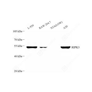
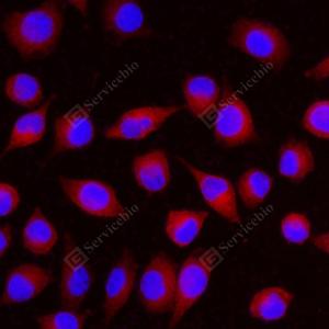
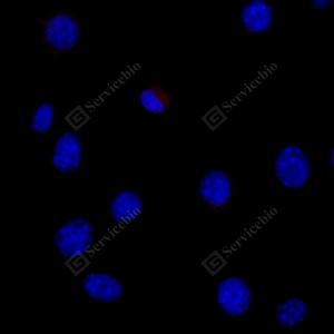
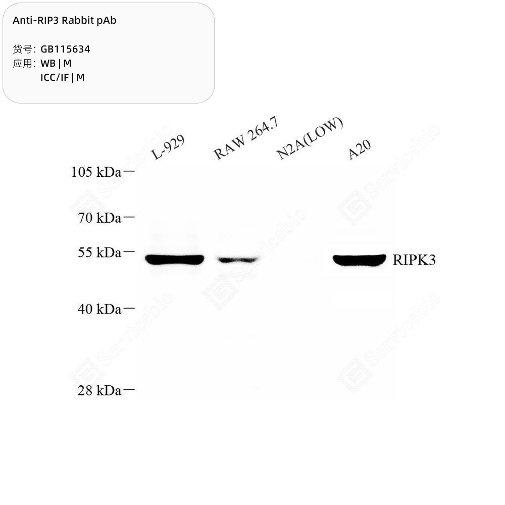
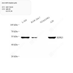
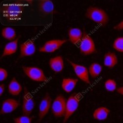
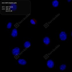
Anti-RIP3 Rabbit pAb

AO-03-GB115634-100
RIP3,Rip3,MPRIP,GB115634,Rho interacting protein 3,GB115634100,M RIP,115634,KIAA0864,RHOIP3,p116Rip,MRIP,Receptor-interacting serine/threonine-protein kinase 3
check En Stock
Hurry! only 10 items left in stock.
429,00 €
HT
Quantité

Product Information


Protein full name

Receptor-interacting serine/threonine-protein kinase 3

Synonyms

Receptor interacting protein 3, RIP 3, RIP like protein kinase 3, RIP3, RIPK3, Rip3 , KIAA0864, M RIP, MPRIP, MRIP, p116Rip, Rho interacting protein 3, RHOIP3

Uniprot ID

Q9QZL0

Immunogen

KLH conjugated Synthetic peptide corresponding to Mouse RIP3

Isotype

IgG

Predicted MW./Observed MW.

53 kDa / 53 kDa

Purity

Affinity purification

Subcellular location

Cytoplasm, cytosol, Nucleus


Applications


Applications Species Dilution Positive Tissue
WB Mouse 1: 500-1: 1000 L-929,RAW 264.7,N2A(low),A20
ICC/IF Mouse 1: 500-1: 1000 L929, NIH-3T3


Background

Serine/threonine-protein kinase that activates necroptosis and apoptosis, two parallel forms of cell death.Necroptosis, a programmed cell death process in response to death-inducing TNF-alpha family members, is triggered by RIPK3 following activation by ZBP1.Activated RIPK3 forms a necrosis-inducing complex and mediates phosphorylation of MLKL, promoting MLKL localization to the plasma membrane and execution of programmed necrosis characterized by calcium influx and plasma membrane damage.In addition to TNF-induced necroptosis, necroptosis can also take place in the nucleus in response to orthomyxoviruses infection: following ZBP1 activation, which senses double-stranded Z-RNA structures, nuclear RIPK3 catalyzes phosphorylation and activation of MLKL, promoting disruption of the nuclear envelope and leakage of cellular DNA into the cytosol.


Images

01W4sZ8nL8eI6OM.jpg

WB analysis of RIP3(GB115634).
Sample: Protein treated by RIPA Lysis Buffer (G2002).
Blocking buffer: 3% Nonfat dry milk (GC310001) in TBST, RT, 1h.
Primary antibody: 1: 500, 4℃ overnight.
Secondary antibody: HRP conjugated Goat Anti-Rabbit lgG (H+L) (GB23303), 1: 5000, RT, 1h.

02pvzM3hbWp1741.jpg

ICC analysis of RIP3(GB115634)red.
Sample: L929, 4% PFA (G1101) 20min.
Blocking buffer: 3% BSA (GC305010) in PBS, RT, 30min.
Primary antibody: 1: 500, 4℃ overnight.
Secondary antibody: Cy3 conjugated Goat Anti-Rabbit IgG (H+L) (GB21303), 1: 300, RT, 1h.

03jpkdK5inumuNF.jpg

ICC analysis of RIP3(GB115634)green.
Sample: NIH-3T3, 4% PFA (G1101) 20min.
Blocking buffer: 3% BSA (GC305010) in PBS, RT, 30min.
Primary antibody: 1: 500, 4℃ overnight.
Secondary antibody: 488 conjugated Goat Anti-Rabbit IgG (H+L) (GB25303), 1: 400, RT, 1h.


Storage


Storage Store at -20℃ for one year. Avoid repeated freeze/thaw cycles.
Storage Buffer PBS with 0.15% ProClin300, 100 μg/mL BSA and 50% glycerol.


NOTE:

1.This product is intended for research only.

2.This product is recommended to dilute with the Primary Antibody Dilution Buffer (G2025).


: AO-03-GB115634-100
: 10 Produits
Hurry! only 10 items left in stock.

Use collapsible tabs for more detailed information that will help customers make a purchasing decision.

Ex: Shipping and return policies, size guides, and other common questions.

  • Paste the label on a flat surface on the package
  • Make sure that both 1D and 2D barcodes are clearly visible
  • Ensure that the label is smooth and isn’t creased or wrinkled
  • Check for any tears, dents, holes or scratches
  • Pack your product tightly, with the right size packaging
  • Ensure both barcodes are on a flat surface of the package