 Français
 English

| ELK.No | ES10135 |
| Product name | RAB9A rabbit pAb |
| Reactivity | Human;Rat;Mouse |
| Applications | WB;ELISA |
| Other name | |
| Size | 50μL |
| Unit price ($) | 148 |
| Human gene ID | 9367 |
| Human Swiss-Prot | P51151 |
| Source | Rabbit |
| Isotype | IgG |
| Target | RAB9A |
| Fields | >>Salmonella infection;>>Measles |
| Gene name | RAB9A RAB9 |
| Protein name | Ras-related protein Rab-9A |
| Human gene link | |
| Human Swiss link | View Human Swiss-Prot |
| Mouse gene ID | |
| Mouse gene link | |
| Mouse Swiss-Prot | Q9R0M6 |
| Mouse Swiss link | View Mouse Swiss-Prot |
| Rat gene ID | |
| Rat gene link | |
| Rat Swiss-Prot | Q99P75 |
| Rat Swiss link | View Rat Swiss-Prot |
| Immunogen | Synthesized peptide derived from human protein . at AA range: 80-160 |
| Specificity | RAB9A Polyclonal Antibody detects endogenous levels of protein. |
| Formulation | Liquid in PBS containing 50% glycerol, and 0.02% sodium azide. |
| Clonality | Polyclonal |
| Dilution | WB 1:500-2000 ELISA 1:5000-20000 |
| Purification | The antibody was affinity-purified from rabbit antiserum by affinity-chromatography using epitope-specific immunogen. |
| Concentration | 1 mg/ml |
| Storage stability | -20°C/1 year |
| Molecular Weight (Da) | |
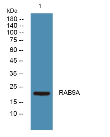
| Observed band (KD) | 22kD |
| Background | function:Involved in the transport of proteins between the endosomes and the trans Golgi network.,similarity:Belongs to the small GTPase superfamily. Rab family., |
| Function | function:Involved in the transport of proteins between the endosomes and the trans Golgi network.,similarity:Belongs to the small GTPase superfamily. Rab family., |
| Subcellular location | Cell membrane ; Lipid-anchor ; Cytoplasmic side . Endoplasmic reticulum membrane . Golgi apparatus membrane . Late endosome . Cytoplasmic vesicle, phagosome membrane ; Lipid-anchor ; Cytoplasmic side . Cytoplasmic vesicle, phagosome . Cytoplasmic vesicle membrane . Melanosome . Colocalizes with OSBPL1A at the late endosome (PubMed:16176980). Recruited to phagosomes containing S.aureus or M.tuberculosis (PubMed:21255211). . |
| Expression | Brain,Epithelium,Muscle, |

Use collapsible tabs for more detailed information that will help customers make a purchasing decision.
Ex: Shipping and return policies, size guides, and other common questions.