 Français
 English
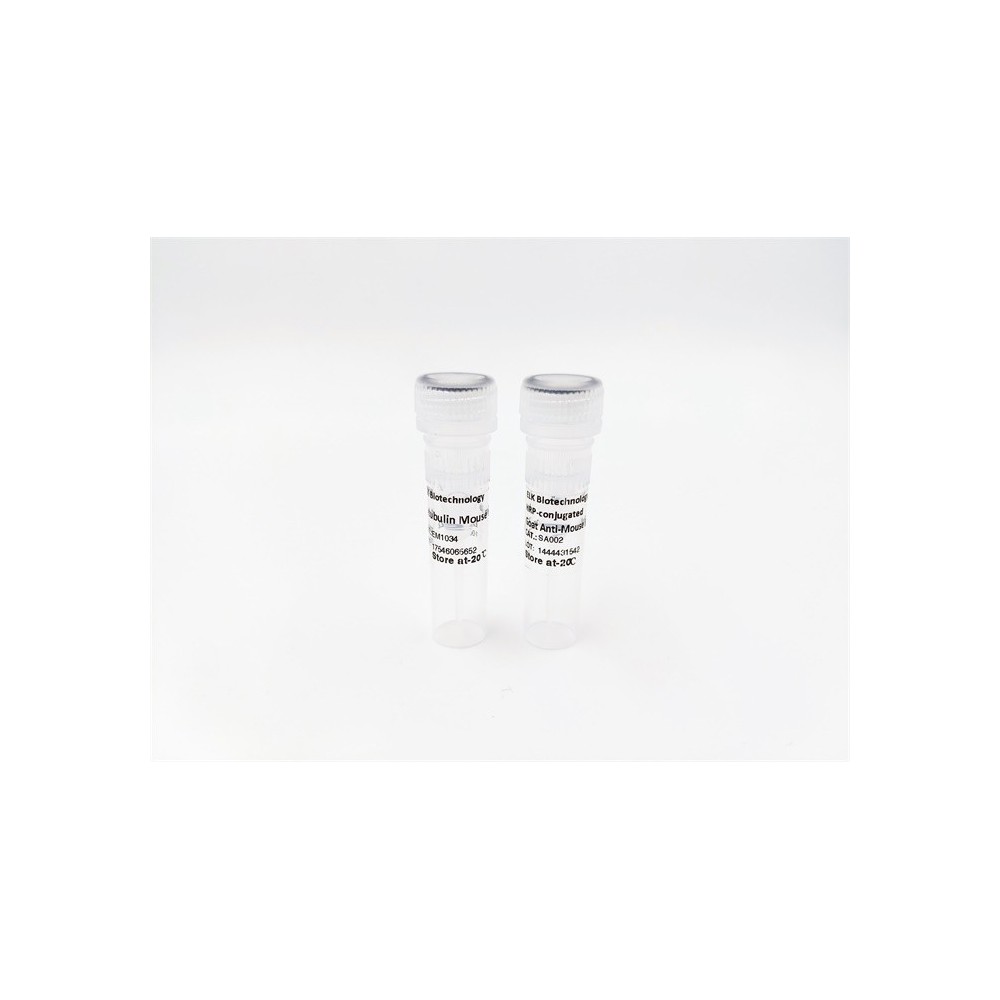

| ELK.No | ES10330 |
| Product name | VAT1 rabbit pAb |
| Reactivity | Human;Rat;Mouse |
| Applications | WB;ELISA |
| Other name | |
| Size | 100μL |
| Unit price ($) | 248 |
| Human gene ID | 10493 |
| Human Swiss-Prot | Q99536 |
| Source | Rabbit |
| Isotype | IgG |
| Target | VAT1 |
| Fields | |
| Gene name | VAT1 |
| Protein name | Synaptic vesicle membrane protein VAT-1 homolog (EC 1.-.-.-) |
| Human gene link | |
| Human Swiss link | View Human Swiss-Prot |
| Mouse gene ID | |
| Mouse gene link | |
| Mouse Swiss-Prot | Q62465 |
| Mouse Swiss link | View Mouse Swiss-Prot |
| Rat gene ID | |
| Rat gene link | |
| Rat Swiss-Prot | Q3MIE4 |
| Rat Swiss link | View Rat Swiss-Prot |
| Immunogen | Synthesized peptide derived from part region of human protein |
| Specificity | VAT1 Polyclonal Antibody detects endogenous levels of protein. |
| Formulation | Liquid in PBS containing 50% glycerol, and 0.02% sodium azide. |
| Clonality | Polyclonal |
| Dilution | WB 1:500-2000 ELISA 1:5000-20000 |
| Purification | The antibody was affinity-purified from rabbit antiserum by affinity-chromatography using epitope-specific immunogen. |
| Concentration | 1 mg/ml |
| Storage stability | -20°C/1 year |
| Molecular Weight (Da) | |
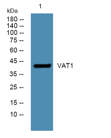
| Observed band (KD) | 43kD |
| Background | vesicle amine transport 1(VAT1) Homo sapiens Synaptic vesicles are responsible for regulating the storage and release of neurotransmitters in the nerve terminal. The protein encoded by this gene is an abundant integral membrane protein of cholinergic synaptic vesicles and is thought to be involved in vesicular transport. It belongs to the quinone oxidoreductase subfamily of zinc-containing alcohol dehydrogenase proteins. [provided by RefSeq, Jul 2008], |
| Function | similarity:Belongs to the zinc-containing alcohol dehydrogenase family. Quinone oxidoreductase subfamily., |
| Subcellular location | Cytoplasm. Mitochondrion outer membrane; Peripheral membrane protein. The majority is localized in the cytoplasm and a small amount is associated with mitochondria. . |
| Expression | Expressed in brain. Also expressed in glioblastoma cells. |

Use collapsible tabs for more detailed information that will help customers make a purchasing decision.
Ex: Shipping and return policies, size guides, and other common questions.