 Français
 English

| ELK.No | ES10548 |
| Product name | MAGAC rabbit pAb |
| Reactivity | Human;Rat;Mouse; |
| Applications | WB;ELISA |
| Other name | |
| Size | 50μL |
| Unit price ($) | 148 |
| Human gene ID | 4111 |
| Human Swiss-Prot | P43365 |
| Source | Rabbit |
| Isotype | IgG |
| Target | MAGAC |
| Fields | |
| Gene name | MAGEA12 MAGE12 |
| Protein name | Melanoma-associated antigen 12 (Cancer/testis antigen 1.12) (CT1.12) (MAGE-12 antigen) (MAGE12F antigen) |
| Human gene link | |
| Human Swiss link | View Human Swiss-Prot |
| Mouse gene ID | |
| Mouse gene link | |
| Mouse Swiss-Prot | |
| Mouse Swiss link | |
| Rat gene ID | |
| Rat gene link | |
| Rat Swiss-Prot | |
| Rat Swiss link | |
| Immunogen | Synthesized peptide derived from human protein . at AA range: 40-120 |
| Specificity | MAGAC Polyclonal Antibody detects endogenous levels of protein. |
| Formulation | Liquid in PBS containing 50% glycerol, and 0.02% sodium azide. |
| Clonality | Polyclonal |
| Dilution | WB 1:500-2000 ELISA 1:5000-20000 |
| Purification | The antibody was affinity-purified from rabbit antiserum by affinity-chromatography using epitope-specific immunogen. |
| Concentration | 1 mg/ml |
| Storage stability | -20°C/1 year |
| Molecular Weight (Da) | |
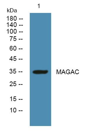
| Observed band (KD) | 34kD |
| Background | MAGE family member A12(MAGEA12) Homo sapiens This gene is closely related to several other genes clustered on chromosome X. These genes may be overexpressed in tumors. Multiple alternatively spliced variants encoding the same protein have been identified. [provided by RefSeq, Jun 2014], |
| Function | function:Not known, though may play a role in tumor transformation or aspects of tumor progression.,similarity:Contains 1 MAGE domain.,tissue specificity:Expressed in many tumors of several types, such as melanoma, head and neck squamous cell carcinoma, lung carcinoma and breast carcinoma, but not in normal tissues except for testes., |
| Subcellular location | |
| Expression | Expressed in many tumors of several types, such as melanoma, head and neck squamous cell carcinoma, lung carcinoma and breast carcinoma, but not in normal tissues except for testes. |

Use collapsible tabs for more detailed information that will help customers make a purchasing decision.
Ex: Shipping and return policies, size guides, and other common questions.