 Français
 English

| ELK.No | ES10561 |
| Product name | MK13 rabbit pAb |
| Reactivity | Human;Mouse;Rat |
| Applications | WB;ELISA |
| Other name | |
| Size | 100μL |
| Unit price ($) | 248 |
| Human gene ID | 5603 |
| Human Swiss-Prot | O15264 |
| Source | Rabbit |
| Isotype | IgG |
| Target | MK13 |
| Fields | >>Endocrine resistance;>>MAPK signaling pathway;>>Rap1 signaling pathway;>>FoxO signaling pathway;>>Sphingolipid signaling pathway;>>Oocyte meiosis;>>Cellular senescence;>>Adrenergic signaling in cardiomyocytes;>>VEGF signaling pathway;>>Osteoclast differentiation;>>Signaling pathways regulating pluripotency of stem cells;>>Platelet activation;>>Neutrophil extracellular trap formation;>>Toll-like receptor signaling pathway;>>NOD-like receptor signaling pathway;>>RIG-I-like receptor signaling pathway;>>C-type lectin receptor signaling pathway;>>IL-17 signaling pathway;>>Th1 and Th2 cell differentiation;>>Th17 cell differentiation;>>T cell receptor signaling pathway;>>Fc epsilon RI signaling pathway;>>TNF signaling pathway;>>Leukocyte transendothelial migration;>>Thermogenesis;>>Neurotrophin signaling pathway;>>Retrograde endocannabinoid signaling;>>Dopaminergic synapse;>>Inflammatory mediator regulation of TRP channels;>>GnRH signaling pathway;>>Progesterone-mediated oocyte maturation;> |
| Gene name | MAPK13 PRKM13 SAPK4 |
| Protein name | Mitogen-activated protein kinase 13 (MAP kinase 13) (MAPK 13) (EC 2.7.11.24) (Mitogen-activated protein kinase p38 delta) (MAP kinase p38 delta) (Stress-activated protein kinase 4) |
| Human gene link | |
| Human Swiss link | View Human Swiss-Prot |
| Mouse gene ID | |
| Mouse gene link | |
| Mouse Swiss-Prot | Q9Z1B7 |
| Mouse Swiss link | View Mouse Swiss-Prot |
| Rat gene ID | |
| Rat gene link | |
| Rat Swiss-Prot | Q9WTY9 |
| Rat Swiss link | View Rat Swiss-Prot |
| Immunogen | Synthesized peptide derived from human protein . at AA range: 200-280 |
| Specificity | MK13 Polyclonal Antibody detects endogenous levels of protein. |
| Formulation | Liquid in PBS containing 50% glycerol, and 0.02% sodium azide. |
| Clonality | Polyclonal |
| Dilution | WB 1:500-2000 ELISA 1:5000-20000 |
| Purification | The antibody was affinity-purified from rabbit antiserum by affinity-chromatography using epitope-specific immunogen. |
| Concentration | 1 mg/ml |
| Storage stability | -20°C/1 year |
| Molecular Weight (Da) | |
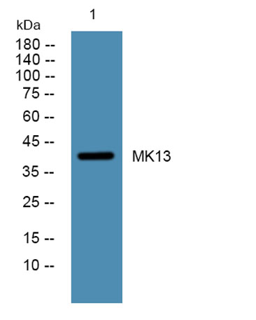
| Observed band (KD) | 40kD |
| Background | This gene encodes a member of the mitogen-activated protein (MAP) kinase family. MAP kinases act as an integration point for multiple biochemical signals, and are involved in a wide variety of cellular processes such as proliferation, differentiation, transcription regulation and development. The encoded protein is a p38 MAP kinase and is activated by proinflammatory cytokines and cellular stress. Substrates of the encoded protein include the transcription factor ATF2 and the microtubule dynamics regulator stathmin. Alternatively spliced transcript variants have been observed for this gene. [provided by RefSeq, Jul 2012], |
| Function | catalytic activity:ATP + a protein = ADP + a phosphoprotein.,cofactor:Magnesium.,domain:The TXY motif contains the threonine and tyrosine residues whose phosphorylation activates the MAP kinases.,enzyme regulation:Activated by phosphorylation on threonine and tyrosine by either of two dual specificity kinases, MAP2K3 and MAP2K6.,function:Responds to activation by environmental stress and pro-inflammatory cytokines by phosphorylating downstream targets. Plays a role in the regulation of protein translation by phosphorylating and inactivating EEF2K.,PTM:Dually phosphorylated on Thr-180 and Tyr-182, which activates the enzyme.,similarity:Belongs to the protein kinase superfamily. CMGC Ser/Thr protein kinase family. MAP kinase subfamily.,similarity:Contains 1 protein kinase domain.,tissue specificity:Expressed in testes, pancreas, small intestine, lung and kidney. Abundant in macrophages, al |
| Subcellular location | intracellular,cytosol, |
| Expression | Expressed in testes, pancreas, small intestine, lung and kidney. Abundant in macrophages, also present in neutrophils, CD4+ T-cells, and endothelial cells. |

Use collapsible tabs for more detailed information that will help customers make a purchasing decision.
Ex: Shipping and return policies, size guides, and other common questions.