 Français
 English

| ELK.No | ES10867 |
| Product name | SLAF7 rabbit pAb |
| Reactivity | Human;Mouse |
| Applications | WB;ELISA |
| Other name | |
| Size | 100μL |
| Unit price ($) | 248 |
| Human gene ID | 57823 |
| Human Swiss-Prot | Q9NQ25 |
| Source | Rabbit |
| Isotype | IgG |
| Target | SLAF7 |
| Fields | |
| Gene name | SLAMF7 CS1 UNQ576/PRO1138 |
| Protein name | SLAM family member 7 (CD2 subset 1) (CD2-like receptor-activating cytotoxic cells) (CRACC) (Membrane protein FOAP-12) (Novel Ly9) (Protein 19A) (CD antigen CD319) |
| Human gene link | |
| Human Swiss link | View Human Swiss-Prot |
| Mouse gene ID | |
| Mouse gene link | |
| Mouse Swiss-Prot | Q8BHK6 |
| Mouse Swiss link | View Mouse Swiss-Prot |
| Rat gene ID | |
| Rat gene link | |
| Rat Swiss-Prot | |
| Rat Swiss link | |
| Immunogen | Synthesized peptide derived from part region of human protein AA range: 41-90 |
| Specificity | SLAF7 Polyclonal Antibody detects endogenous levels of protein. |
| Formulation | Liquid in PBS containing 50% glycerol, and 0.02% sodium azide. |
| Clonality | Polyclonal |
| Dilution | WB 1:500-2000 ELISA 1:5000-20000 |
| Purification | The antibody was affinity-purified from rabbit antiserum by affinity-chromatography using epitope-specific immunogen. |
| Concentration | 1 mg/ml |
| Storage stability | -20°C/1 year |
| Molecular Weight (Da) | |
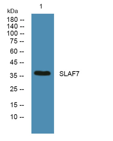
| Observed band (KD) | 36kD |
| Background | function:Isoform 1 mediates NK cell activation through a SAP-independent extracellular signal-regulated ERK-mediated pathway. May play a role in lymphocyte adhesion. Isoform 3 does not mediate any activation. SAP can bind the cytoplasmic tail of isoform 1 when phosphorylated in the presence of Fyn (in vitro).,similarity:Contains 1 Ig-like C2-type (immunoglobulin-like) domain.,tissue specificity:Expressed in spleen, lymph node, peripheral blood leukocytes, bone marrow, small intestine, stomach, appendix, lung and trachea. Expression was detected in NK cells, activated B-cells, NK-cell line but not in promyelocytic, B-, or T-cell lines. The isoform 3 is expressed at much lower level tha isoform 1., |
| Function | function:Isoform 1 mediates NK cell activation through a SAP-independent extracellular signal-regulated ERK-mediated pathway. May play a role in lymphocyte adhesion. Isoform 3 does not mediate any activation. SAP can bind the cytoplasmic tail of isoform 1 when phosphorylated in the presence of Fyn (in vitro).,similarity:Contains 1 Ig-like C2-type (immunoglobulin-like) domain.,tissue specificity:Expressed in spleen, lymph node, peripheral blood leukocytes, bone marrow, small intestine, stomach, appendix, lung and trachea. Expression was detected in NK cells, activated B-cells, NK-cell line but not in promyelocytic, B-, or T-cell lines. The isoform 3 is expressed at much lower level tha isoform 1., |
| Subcellular location | Membrane; Single-pass type I membrane protein. |
| Expression | Expressed in spleen, lymph node, peripheral blood leukocytes, bone marrow, small intestine, stomach, appendix, lung and trachea. Expression was detected in NK cells, activated B-cells, NK-cell line but not in promyelocytic, B-, or T-cell lines. Expressed in monocytes. Isoform 3 is expressed at much lower level than isoform 1. |

Use collapsible tabs for more detailed information that will help customers make a purchasing decision.
Ex: Shipping and return policies, size guides, and other common questions.