 Français
 English

| ELK.No | ES11111 |
| Product name | Iba1 rabbit pAb |
| Reactivity | Human;Rat;Mouse |
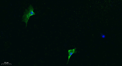
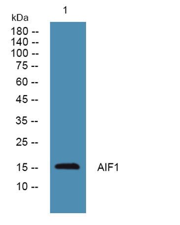
| Applications | WB;IF;ELISA |
| Other name | |
| Size | 100μL |
| Unit price ($) | 248 |
| Human gene ID | 199 |
| Human Swiss-Prot | P55008 |
| Source | Rabbit |
| Isotype | IgG |
| Target | AIF1 |
| Fields | |
| Gene name | AIF1 G1 IBA1 |
| Protein name | Allograft inflammatory factor 1 (AIF-1) (Ionized calcium-binding adapter molecule 1) (Protein G1) |
| Human gene link | |
| Human Swiss link | View Human Swiss-Prot |
| Mouse gene ID | |
| Mouse gene link | |
| Mouse Swiss-Prot | O70200 |
| Mouse Swiss link | View Mouse Swiss-Prot |
| Rat gene ID | |
| Rat gene link | |
| Rat Swiss-Prot | P55009 |
| Rat Swiss link | View Rat Swiss-Prot |
| Immunogen | Synthesized peptide derived from part region of human protein |
| Specificity | AIF1 Polyclonal Antibody detects endogenous levels of protein. |
| Formulation | Liquid in PBS containing 50% glycerol, and 0.02% sodium azide. |
| Clonality | Polyclonal |
| Dilution | Western Blot: 1/500 - 1/2000. ELISA: 1/5000. IF 1:100-300 Not yet tested in other applications. |
| Purification | The antibody was affinity-purified from rabbit antiserum by affinity-chromatography using epitope-specific immunogen. |
| Concentration | 1 mg/ml |
| Storage stability | -20°C/1 year |
| Molecular Weight (Da) | |
| Observed band (KD) | 16kD |
| Background | This gene encodes a protein that binds actin and calcium. This gene is induced by cytokines and interferon and may promote macrophage activation and growth of vascular smooth muscle cells and T-lymphocytes. Polymorphisms in this gene may be associated with systemic sclerosis. Alternative splicing results in multiple transcript variants, but the full-length and coding nature of some of these variants is not certain. [provided by RefSeq, Jan 2016], |
| Function | function:May play a role in macrophage activation and function.,PTM:Phosphorylated on serine residues.,similarity:Contains 2 EF-hand domains., |
| Subcellular location | Cytoplasm, cytoskeleton . Cell projection, ruffle membrane ; Peripheral membrane protein ; Cytoplasmic side . Cell projection, phagocytic cup . Associated with the actin cytoskeleton at membrane ruffles and at sites of phagocytosis. . |
| Expression | Detected in T-lymphocytes and peripheral blood mononuclear cells. |

Use collapsible tabs for more detailed information that will help customers make a purchasing decision.
Ex: Shipping and return policies, size guides, and other common questions.