 Français
 English
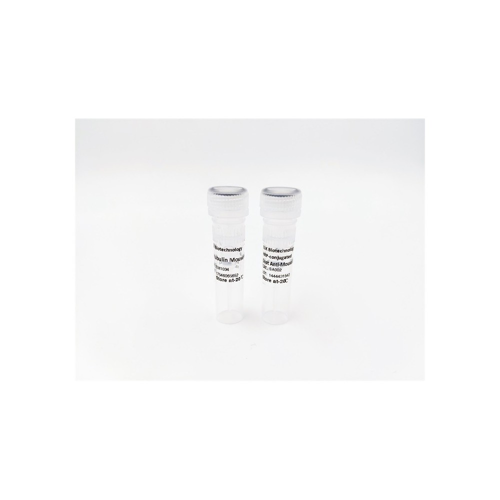

| ELK.No | ES11400 |
| Product name | SCTM1 rabbit pAb |
| Reactivity | Human;Rat;Mouse; |
| Applications | WB;ELISA |
| Other name | |
| Size | 100μL |
| Unit price ($) | 248 |
| Human gene ID | 6398 |
| Human Swiss-Prot | Q8WVN6 |
| Source | Rabbit |
| Isotype | IgG |
| Target | SCTM1 |
| Fields | |
| Gene name | SECTM1 K12 |
| Protein name | Secreted and transmembrane protein 1 (Protein K-12) |
| Human gene link | |
| Human Swiss link | View Human Swiss-Prot |
| Mouse gene ID | |
| Mouse gene link | |
| Mouse Swiss-Prot | |
| Mouse Swiss link | |
| Rat gene ID | |
| Rat gene link | |
| Rat Swiss-Prot | |
| Rat Swiss link | |
| Immunogen | Synthesized peptide derived from human protein . at AA range: 71-120 |
| Specificity | SCTM1 Polyclonal Antibody detects endogenous levels of protein. |
| Formulation | Liquid in PBS containing 50% glycerol, and 0.02% sodium azide. |
| Clonality | Polyclonal |
| Dilution | WB 1:500-2000 ELISA 1:5000-20000 |
| Purification | The antibody was affinity-purified from rabbit antiserum by affinity-chromatography using epitope-specific immunogen. |
| Concentration | 1 mg/ml |
| Storage stability | -20°C/1 year |
| Molecular Weight (Da) | |
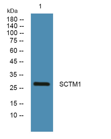
| Observed band (KD) | 27kD |
| Background | This gene encodes a transmembrane and secreted protein with characteristics of a type 1a transmembrane protein. It is found in a perinuclear Golgi-like pattern and thought to be involved in hematopoietic and/or immune system processes. [provided by RefSeq, Jul 2008], |
| Function | subunit:Interacts with CD7.,tissue specificity:Detected at the highest levels in peripheral blood leukocytes and breast cancer cell lines. Found in leukocytes of the myeloid lineage, with the strongest expression observed in granulocytes and no detectable expression in lymphocytes., |
| Subcellular location | Cell membrane ; Single-pass type I membrane protein . Secreted. |
| Expression | Detected at the highest levels in peripheral blood leukocytes and breast cancer cell lines. Found in leukocytes of the myeloid lineage, with the strongest expression observed in granulocytes and no detectable expression in lymphocytes. Expressed in thymic epithelial cells and fibroblasts. |

Use collapsible tabs for more detailed information that will help customers make a purchasing decision.
Ex: Shipping and return policies, size guides, and other common questions.