 Français
 English

| ELK.No | ES11500 |
| Product name | HCAR1 rabbit pAb |
| Reactivity | Human;Rat;Mouse; |
| Applications | WB;ELISA |
| Other name | |
| Size | 50μL |
| Unit price ($) | 148 |
| Human gene ID | 27198 |
| Human Swiss-Prot | Q9BXC0 |
| Source | Rabbit |
| Isotype | IgG |
| Target | HCAR1 |
| Fields | >>cAMP signaling pathway |
| Gene name | HCAR1 GPR104 GPR81 HCA1 FKSG80 |
| Protein name | Hydroxycarboxylic acid receptor 1 (G-protein coupled receptor 104) (G-protein coupled receptor 81) |
| Human gene link | |
| Human Swiss link | View Human Swiss-Prot |
| Mouse gene ID | |
| Mouse gene link | |
| Mouse Swiss-Prot | Q8C131 |
| Mouse Swiss link | View Mouse Swiss-Prot |
| Rat gene ID | |
| Rat gene link | |
| Rat Swiss-Prot | |
| Rat Swiss link | |
| Immunogen | Synthesized peptide derived from part region of human protein |
| Specificity | HCAR1 Polyclonal Antibody detects endogenous levels of protein. |
| Formulation | Liquid in PBS containing 50% glycerol, and 0.02% sodium azide. |
| Clonality | Polyclonal |
| Dilution | WB 1:500-2000 ELISA 1:5000-20000 |
| Purification | The antibody was affinity-purified from rabbit antiserum by affinity-chromatography using epitope-specific immunogen. |
| Concentration | 1 mg/ml |
| Storage stability | -20°C/1 year |
| Molecular Weight (Da) | |
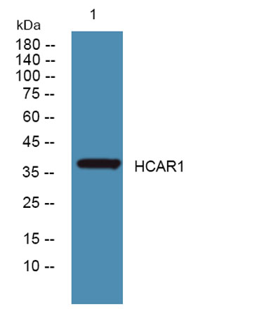
| Observed band (KD) | 38kD |
| Background | G protein-coupled receptors (GPCRs, or GPRs), such as GPR81, contain 7 transmembrane domains and transduce extracellular signals through heterotrimeric G proteins.[supplied by OMIM, Feb 2005], |
| Function | function:Orphan receptor.,similarity:Belongs to the G-protein coupled receptor 1 family.,tissue specificity:Expressed in pituitary tissue. Not detected in frontal, temporal and occipital lobes of the cortex, basal forebrain, caudate nucleus, nucleus accumbens, and hippocampus., |
| Subcellular location | Cell membrane; Multi-pass membrane protein. |
| Expression | Expressed abundantly in brown and white fat. It also detectable at lower levels in liver, kidney, skeletal muscle, brain and pituitary. Not detected in frontal, temporal and occipital lobes of the cortex, basal forebrain, caudate nucleus, nucleus accumbens and hippocampus. |

Use collapsible tabs for more detailed information that will help customers make a purchasing decision.
Ex: Shipping and return policies, size guides, and other common questions.