 Français
 English
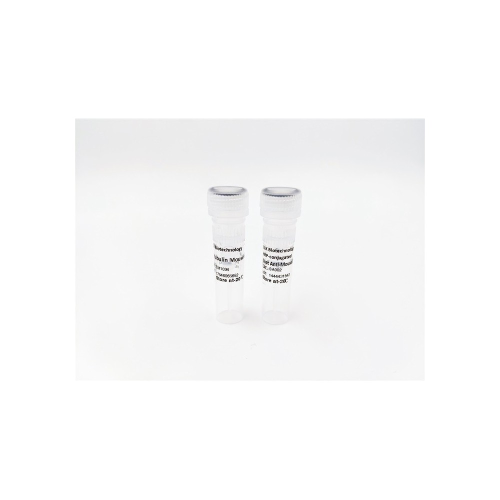

| ELK.No | ES11532 |
| Product name | OPSB rabbit pAb |
| Reactivity | Human;Rat;Mouse; |
| Applications | WB;ELISA |
| Other name | |
| Size | 50μL |
| Unit price ($) | 148 |
| Human gene ID | 611 |
| Human Swiss-Prot | P03999 |
| Source | Rabbit |
| Isotype | IgG |
| Target | OPSB |
| Fields | |
| Gene name | OPN1SW BCP |
| Protein name | Short-wave-sensitive opsin 1 (Blue cone photoreceptor pigment) (Blue-sensitive opsin) (BOP) |
| Human gene link | |
| Human Swiss link | View Human Swiss-Prot |
| Mouse gene ID | |
| Mouse gene link | |
| Mouse Swiss-Prot | P51491 |
| Mouse Swiss link | View Mouse Swiss-Prot |
| Rat gene ID | |
| Rat gene link | |
| Rat Swiss-Prot | Q63652 |
| Rat Swiss link | View Rat Swiss-Prot |
| Immunogen | Synthesized peptide derived from part region of human protein |
| Specificity | OPSB Polyclonal Antibody detects endogenous levels of protein. |
| Formulation | Liquid in PBS containing 50% glycerol, and 0.02% sodium azide. |
| Clonality | Polyclonal |
| Dilution | WB 1:500-2000 ELISA 1:5000-20000 |
| Purification | The antibody was affinity-purified from rabbit antiserum by affinity-chromatography using epitope-specific immunogen. |
| Concentration | 1 mg/ml |
| Storage stability | -20°C/1 year |
| Molecular Weight (Da) | |
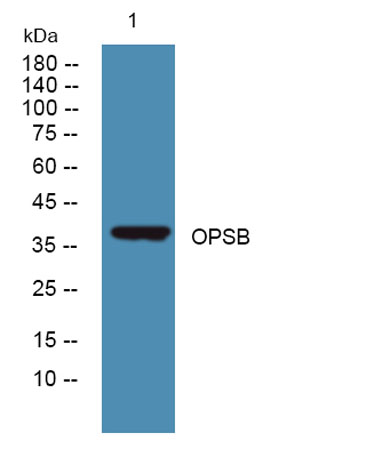
| Observed band (KD) | 38kD |
| Background | This gene belongs to the G-protein coupled receptor 1 family, opsin subfamily. It encodes the blue cone pigment gene which is one of three types of cone photoreceptors responsible for normal color vision. Defects in this gene are the cause of tritan color blindness (tritanopia). Affected individuals lack blue and yellow sensory mechanisms while retaining those for red and green. Defective blue vision is characteristic. [provided by RefSeq, Jul 2008], |
| Function | disease:Defects in OPN1SW are the cause of tritan color blindness (tritanopia) [MIM:190900].,function:Visual pigments are the light-absorbing molecules that mediate vision. They consist of an apoprotein, opsin, covalently linked to cis-retinal.,online information:Retina International's Scientific Newsletter,PTM:Phosphorylated on some or all of the serine and threonine residues present in the C-terminal region.,similarity:Belongs to the G-protein coupled receptor 1 family.,similarity:Belongs to the G-protein coupled receptor 1 family. Opsin subfamily.,tissue specificity:The three color pigments are found in the cone photoreceptor cells., |
| Subcellular location | Cell membrane ; Multi-pass membrane protein . Photoreceptor inner segment . Cell projection, cilium, photoreceptor outer segment . Cytoplasm, perinuclear region . |
| Expression | The three color pigments are found in the cone photoreceptor cells (PubMed:2937147). Expressed throughout the epidermis and dermis, primarily in the stratum granulosum in the facial and abdominal skin (at protein level) (PubMed:30168605). Expressed in dermal fibroblasts (at protein level) (PubMed:31380578). Expressed in melanocytes (at protein level) (PubMed:31730232). |

Use collapsible tabs for more detailed information that will help customers make a purchasing decision.
Ex: Shipping and return policies, size guides, and other common questions.