 Français
 English
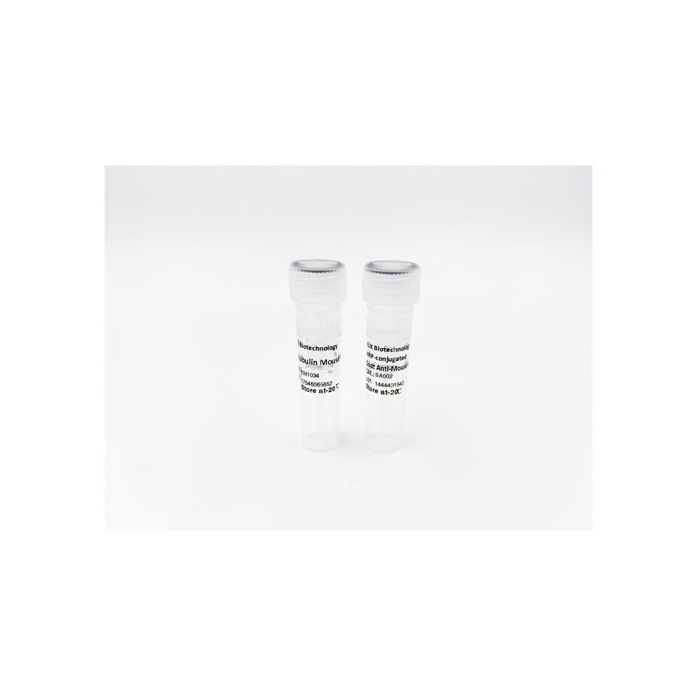

| ELK.No | ES12323 |
| Product name | WDR33 rabbit pAb |
| Reactivity | Human; Mouse |
| Applications | WB |
| Other name | |
| Size | 50μL |
| Unit price ($) | 148 |
| Human gene ID | 55339 |
| Human Swiss-Prot | Q9C0J8 |
| Source | Rabbit |
| Isotype | IgG |
| Target | WDR33 |
| Fields | >>mRNA surveillance pathway |
| Gene name | WDR33 WDC146 |
| Protein name | WDR33 |
| Human gene link | View Human Gene |
| Human Swiss link | View Human Swiss-Prot |
| Mouse gene ID | 74320 |
| Mouse gene link | View Mouse Gene |
| Mouse Swiss-Prot | Q8K4P0 |
| Mouse Swiss link | View Mouse Swiss-Prot |
| Rat gene ID | |
| Rat gene link | |
| Rat Swiss-Prot | |
| Rat Swiss link | |
| Immunogen | Synthesized peptide derived from human WDR33 AA range: 319-369 |
| Specificity | This antibody detects endogenous levels of WDR33 at Human/Mouse |
| Formulation | Liquid in PBS containing 50% glycerol, 0.5% BSA and 0.02% sodium azide. |
| Clonality | Polyclonal |
| Dilution | WB 1:500-2000 |
| Purification | The antibody was affinity-purified from rabbit serum by affinity-chromatography using specific immunogen. |
| Concentration | 1 mg/ml |
| Storage stability | -20°C/1 year |
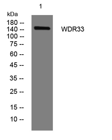
| Molecular Weight (Da) | 147kD |
| Observed band (KD) | |
| Background | This gene encodes a member of the WD repeat protein family. WD repeats are minimally conserved regions of approximately 40 amino acids typically bracketed by gly-his and trp-asp (GH-WD), which may facilitate formation of heterotrimeric or multiprotein complexes. Members of this family are involved in a variety of cellular processes, including cell cycle progression, signal transduction, apoptosis, and gene regulation. This gene is highly expressed in testis and the protein is localized to the nucleus. This gene may play important roles in the mechanisms of cytodifferentiation and/or DNA recombination. Multiple alternatively spliced transcript variants encoding distinct isoforms have been found for this gene. [provided by RefSeq, Jul 2008], |
| Function | similarity:Contains 1 collagen-like domain.,similarity:Contains 7 WD repeats.,tissue specificity:Most highly expressed in testis., |
| Subcellular location | Nucleus . |
| Expression | Most highly expressed in testis. |

Use collapsible tabs for more detailed information that will help customers make a purchasing decision.
Ex: Shipping and return policies, size guides, and other common questions.