 Français
 English
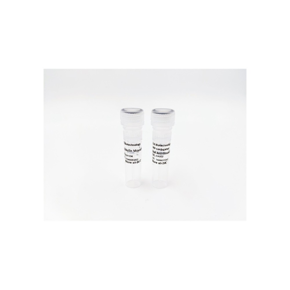

| ELK.No | ES12415 |
| Product name | URGCP rabbit pAb |
| Reactivity | Human; Mouse |
| Applications | WB |
| Other name | |
| Size | 50μL |
| Unit price ($) | 148 |
| Human gene ID | 55665 |
| Human Swiss-Prot | Q8TCY9 |
| Source | Rabbit |
| Isotype | IgG |
| Target | URGCP |
| Fields | |
| Gene name | URGCP KIAA1507 URG4 |
| Protein name | URGCP |
| Human gene link | View Human Gene |
| Human Swiss link | View Human Swiss-Prot |
| Mouse gene ID | 72046 |
| Mouse gene link | View Mouse Gene |
| Mouse Swiss-Prot | Q5NCI0 |
| Mouse Swiss link | View Mouse Swiss-Prot |
| Rat gene ID | |
| Rat gene link | |
| Rat Swiss-Prot | |
| Rat Swiss link | |
| Immunogen | Synthesized peptide derived from human URGCP AA range: 345-395 |
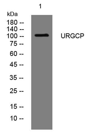
| Specificity | This antibody detects endogenous levels of URGCP at Human/Mouse |
| Formulation | Liquid in PBS containing 50% glycerol, 0.5% BSA and 0.02% sodium azide. |
| Clonality | Polyclonal |
| Dilution | WB 1:500-2000 |
| Purification | The antibody was affinity-purified from rabbit serum by affinity-chromatography using specific immunogen. |
| Concentration | 1 mg/ml |
| Storage stability | -20°C/1 year |
| Molecular Weight (Da) | 102kD |
| Observed band (KD) | |
| Background | URG4 is upregulated in the presence of hepatitis B virus (HBV)-encoded X antigen (HBxAg) and may contribute to the development of hepatocellular carcinoma by promoting hepatocellular growth and survival (Tufan et al., 2002 [PubMed 12082552]).[supplied by OMIM, Mar 2008], |
| Function | function:May be involved in cell cycle progression through the regulation of cyclin D1 expression. May participate in the development of hepatocellular carcinoma (HCC) by promoting hepatocellular growth and survival. May play an important role in development of gastric cancer.,induction:By HBxAg. Up-regulated in gastric cancer tissues and also in gastric cancer cell lines (at protein level).,sequence caution:Translation N-terminally extended.,subcellular location:In epithelial cells localized predominantly in the cytoplasm and occasionally in nuclei.,tissue specificity:Strongly expressed in hepatitis B-infected liver and in HCC cells. Also highly expressed in well-differentiated gastric cancer tissues and various gastric cancer cell lines., |
| Subcellular location | Cytoplasm . Nucleus . In epithelial cells localized predominantly in the cytoplasm and occasionally in nuclei. |
| Expression | Strongly expressed in hepatitis B virus-infected liver and in HCC cells. Also highly expressed in well-differentiated gastric cancer tissues and various gastric cancer cell lines. |

Use collapsible tabs for more detailed information that will help customers make a purchasing decision.
Ex: Shipping and return policies, size guides, and other common questions.