 Français
 English
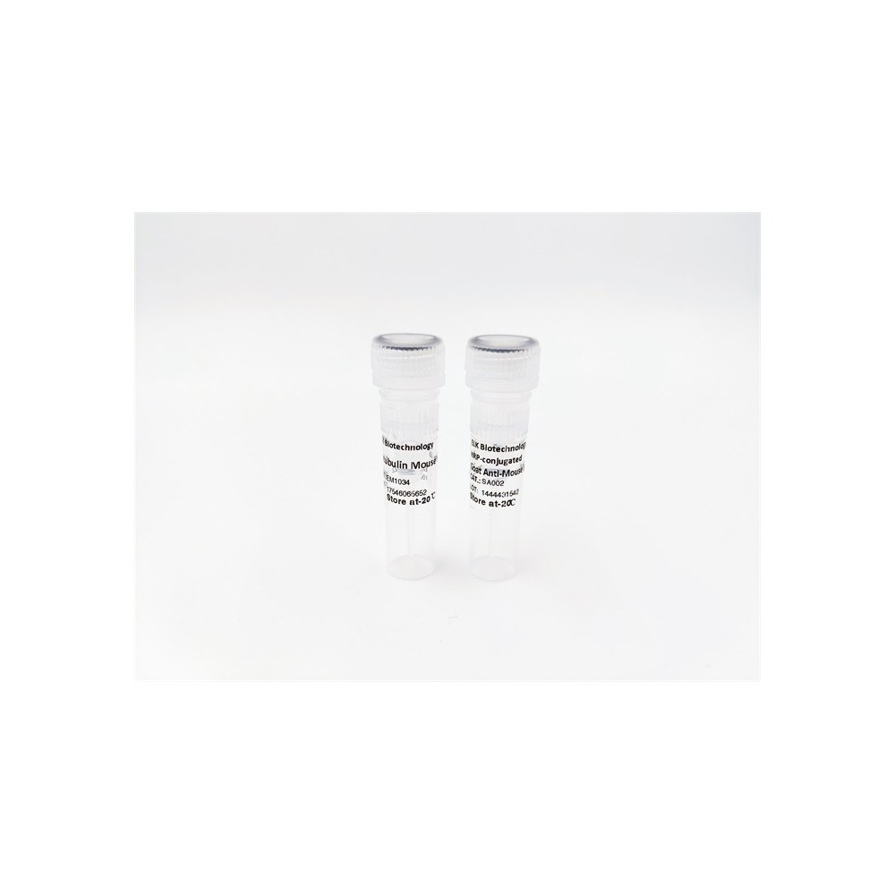

| ELK.No | ES12626 |
| Product name | TMX1 rabbit pAb |
| Reactivity | Human; Mouse |
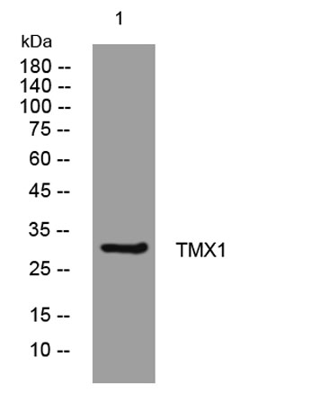
| Applications | WB |
| Other name | |
| Size | 50μL |
| Unit price ($) | 148 |
| Human gene ID | 81542 |
| Human Swiss-Prot | Q9H3N1 |
| Source | Rabbit |
| Isotype | IgG |
| Target | TMX1 |
| Fields | |
| Gene name | TMX1 TMX TXNDC TXNDC1 PSEC0085 UNQ235/PRO268 |
| Protein name | TMX1 |
| Human gene link | View Human Gene |
| Human Swiss link | View Human Swiss-Prot |
| Mouse gene ID | 72736 |
| Mouse gene link | View Mouse Gene |
| Mouse Swiss-Prot | Q8VBT0 |
| Mouse Swiss link | View Mouse Swiss-Prot |
| Rat gene ID | |
| Rat gene link | |
| Rat Swiss-Prot | |
| Rat Swiss link | |
| Immunogen | Synthesized peptide derived from human TMX1 AA range: 223-273 |
| Specificity | This antibody detects endogenous levels of TMX1 at Human/Mouse |
| Formulation | Liquid in PBS containing 50% glycerol, 0.5% BSA and 0.02% sodium azide. |
| Clonality | Polyclonal |
| Dilution | WB 1:500-2000 |
| Purification | The antibody was affinity-purified from rabbit serum by affinity-chromatography using specific immunogen. |
| Concentration | 1 mg/ml |
| Storage stability | -20°C/1 year |
| Molecular Weight (Da) | 31kD |
| Observed band (KD) | |
| Background | TXNDC1 is a thioredoxin (TXN; see MIM 187700)-related protein with disulfide reductase activity (Matsuo et al., 2001 [PubMed 11152479]).[supplied by OMIM, Mar 2008], |
| Function | function:May participate in various redox reactions through the reversible oxidation of its active center dithiol to a disulfide and catalyze dithiol-disulfide exchange reactions.,similarity:Contains 1 thioredoxin domain.,subcellular location:Predominantly found in the endoplasmic reticulum.,tissue specificity:Ubiquitous. Highly expressed in kidney, liver, placenta and lung., |
| Subcellular location | Membrane ; Single-pass type I membrane protein . Endoplasmic reticulum membrane ; Single-pass type I membrane protein . Predominantly found in the endoplasmic reticulum. . |
| Expression | Ubiquitous. Highly expressed in kidney, liver, placenta and lung. |

Use collapsible tabs for more detailed information that will help customers make a purchasing decision.
Ex: Shipping and return policies, size guides, and other common questions.