 Français
 English

| ELK.No | ES12693 |
| Product name | TINAG rabbit pAb |
| Reactivity | Human;Rat;Mouse; |
| Applications | WB;IHC |
| Other name | |
| Size | 50μL |
| Unit price ($) | 148 |
| Human gene ID | 27283 |
| Human Swiss-Prot | Q9UJW2 |
| Source | Rabbit |
| Isotype | IgG |
| Target | TINAG |
| Fields | |
| Gene name | TINAG |
| Protein name | TINAG |
| Human gene link | View Human Gene |
| Human Swiss link | View Human Swiss-Prot |
| Mouse gene ID | |
| Mouse gene link | |
| Mouse Swiss-Prot | |
| Mouse Swiss link | |
| Rat gene ID | |
| Rat gene link | |
| Rat Swiss-Prot | |
| Rat Swiss link | |
| Immunogen | Synthesized peptide derived from human TINAG AA range: 80-130 |
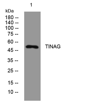
| Specificity | This antibody detects endogenous levels of TINAG at Human |
| Formulation | Liquid in PBS containing 50% glycerol, 0.5% BSA and 0.02% sodium azide. |
| Clonality | Polyclonal |
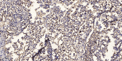
| Dilution | WB 1:500-2000;IHC-p 1:50-300 |
| Purification | The antibody was affinity-purified from rabbit serum by affinity-chromatography using specific immunogen. |
| Concentration | 1 mg/ml |
| Storage stability | -20°C/1 year |
| Molecular Weight (Da) | 52kD |
| Observed band (KD) | |
| Background | This gene encodes a glycoprotein that is restricted within the kidney to the basement membranes underlying the epithelium of Bowman's capsule and proximal and distal tubules. Autoantibodies against this protein are found in sera of patients with tubulointerstital nephritis, membranous nephropathy and anti-glomerular basement membrane nephritis. Ontogeny studies suggest that the expression of this antigen is developmentally regulated in a precise spatial and temporal pattern throughout nephrogenesis. [provided by RefSeq, Nov 2011], |
| Function | developmental stage:Initially observed in the Bowman capsule during early glomerular capillary loop formation in the kidney. In more developmentally mature glomeruli, following transition from early to mid-capillary loop stage, expression is higher in the proximal tubular basement membrane than in the distal basement membrane and Bowman capsule.,disease:Antibodies against TINAG are found in sera of patients with tubulointerstitial nephritis, a rare autoimmune disorder that causes acute and chronic renal injury.,function:Mediates adhesion of proximal tubule epithelial cells via integrins alpha3-beta1 and alphaV-beta3.,PTM:It has been suggested that the active SMB domain may be permitted considerable disulfide bond heterogeneity or variability, thus 2 alternate disulfide patterns based on 3D structures are described with 1 disulfide bond conserved in both.,similarity:Belongs to the peptida |
| Subcellular location | Secreted, extracellular space, extracellular matrix, basement membrane . |
| Expression | Expressed in the kidney cortex, small intestine and cornea. |

Use collapsible tabs for more detailed information that will help customers make a purchasing decision.
Ex: Shipping and return policies, size guides, and other common questions.