 Français
 English

| ELK.No | ES12906 |
| Product name | STBD1 rabbit pAb |
| Reactivity | Human; Mouse;Rat |
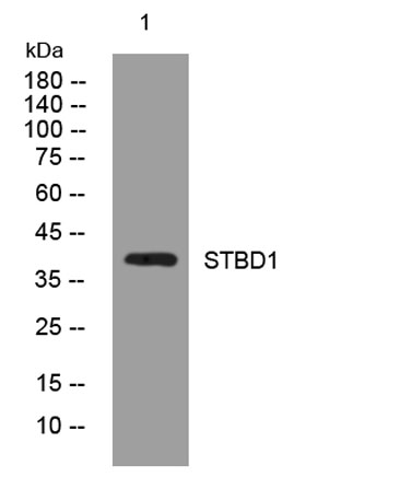
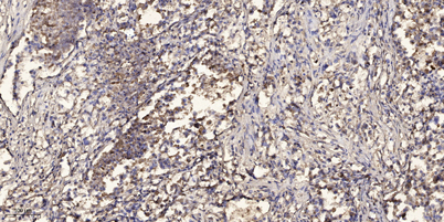
| Applications | WB;ELISA;IHC |
| Other name | |
| Size | 100μL |
| Unit price ($) | 248 |
| Human gene ID | 8987 |
| Human Swiss-Prot | O95210 |
| Source | Rabbit |
| Isotype | IgG |
| Target | STBD1 |
| Fields | |
| Gene name | STBD1 GENX-3414 |
| Protein name | STBD1 |
| Human gene link | View Human Gene |
| Human Swiss link | View Human Swiss-Prot |
| Mouse gene ID | 52331 |
| Mouse gene link | View Mouse Gene |
| Mouse Swiss-Prot | Q8C7E7 |
| Mouse Swiss link | View Mouse Swiss-Prot |
| Rat gene ID | 305234 |
| Rat gene link | View Rat Gene |
| Rat Swiss-Prot | Q5FVN1 |
| Rat Swiss link | View Rat Swiss-Prot |
| Immunogen | Synthesized peptide derived from human STBD1 AA range: 107-157 |
| Specificity | This antibody detects endogenous levels of STBD1 at Human/Mouse/Rat |
| Formulation | Liquid in PBS containing 50% glycerol, 0.5% BSA and 0.02% sodium azide. |
| Clonality | Polyclonal |
| Dilution | WB 1:500-2000;IHC-p 1:50-300; ELISA 2000-20000 |
| Purification | The antibody was affinity-purified from rabbit serum by affinity-chromatography using specific immunogen. |
| Concentration | 1 mg/ml |
| Storage stability | -20°C/1 year |
| Molecular Weight (Da) | 39kD |
| Observed band (KD) | |
| Background | |
| Function | similarity:Contains 1 CBM20 (carbohydrate binding type-20) domain.,subcellular location:Distributed in the transverse tubules and/or near the junctional sarcoplasmic reticulum.,tissue specificity:Expressed at high level in skeletal and cardiac muscles. Moderately expressed in liver and placenta. No expression is found in pancreas, kidney or lung. Present in skeletal muscle, heart and placenta (at protein level)., |
| Subcellular location | Preautophagosomal structure membrane ; Single-pass type III membrane protein . Endoplasmic reticulum membrane ; Single-pass type III membrane protein . Cell membrane, sarcolemma, T-tubule . Also detected near the junctional sarcoplasmic reticulum (PubMed:9794794). Concentrates at perinuclear structures (PubMed:21893048). . |
| Expression | Expressed at high level in skeletal and cardiac muscles. Moderately expressed in liver and placenta. No expression is found in pancreas, kidney or lung. Present in skeletal muscle, heart and placenta (at protein level). |

Use collapsible tabs for more detailed information that will help customers make a purchasing decision.
Ex: Shipping and return policies, size guides, and other common questions.