 Français
 English

| ELK.No | ES12940 |
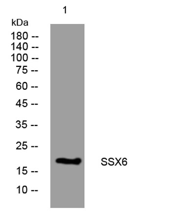
| Product name | SSX6 rabbit pAb |
| Reactivity | Human;Rat;Mouse; |
| Applications | WB |
| Other name | |
| Size | 50μL |
| Unit price ($) | 148 |
| Human gene ID | |
| Human Swiss-Prot | Q7RTT6 |
| Source | Rabbit |
| Isotype | IgG |
| Target | SSX6 |
| Fields | |
| Gene name | SSX6 |
| Protein name | SSX6 |
| Human gene link | |
| Human Swiss link | View Human Swiss-Prot |
| Mouse gene ID | |
| Mouse gene link | |
| Mouse Swiss-Prot | |
| Mouse Swiss link | |
| Rat gene ID | |
| Rat gene link | |
| Rat Swiss-Prot | |
| Rat Swiss link | |
| Immunogen | Synthesized peptide derived from human SSX6 AA range: 119-169 |
| Specificity | This antibody detects endogenous levels of SSX6 at Human |
| Formulation | Liquid in PBS containing 50% glycerol, 0.5% BSA and 0.02% sodium azide. |
| Clonality | Polyclonal |
| Dilution | WB 1:500-2000 |
| Purification | The antibody was affinity-purified from rabbit serum by affinity-chromatography using specific immunogen. |
| Concentration | 1 mg/ml |
| Storage stability | -20°C/1 year |
| Molecular Weight (Da) | 21kD |
| Observed band (KD) | |
| Background | This gene belongs to the family of highly homologous synovial sarcoma X (SSX) breakpoint proteins. These proteins may function as transcriptional repressors. They are also capable of eliciting spontaneously humoral and cellular immune responses in cancer patients, and are potentially useful targets in cancer vaccine-based immunotherapy. SSX1, SSX2 and SSX4 genes have been involved in the t(X;18) translocation characteristically found in all synovial sarcomas. This gene is classified as a pseudogene because a splice donor in the 3' UTR has changed compared to other family members, rendering the transcript a candidate for nonsense-mediated mRNA decay (NMD). [provided by RefSeq, Aug 2009], |
| Function | function:Could act as a modulator of transcription.,similarity:Belongs to the SSX family.,similarity:Contains 1 KRAB-related domain.,tissue specificity:Not detected in any normal tissues. Expressed in a melanoma cell line., |
| Subcellular location | nucleus, |
| Expression | Not detected in any normal tissues. Expressed in a melanoma cell line. |

Use collapsible tabs for more detailed information that will help customers make a purchasing decision.
Ex: Shipping and return policies, size guides, and other common questions.