 Français
 English
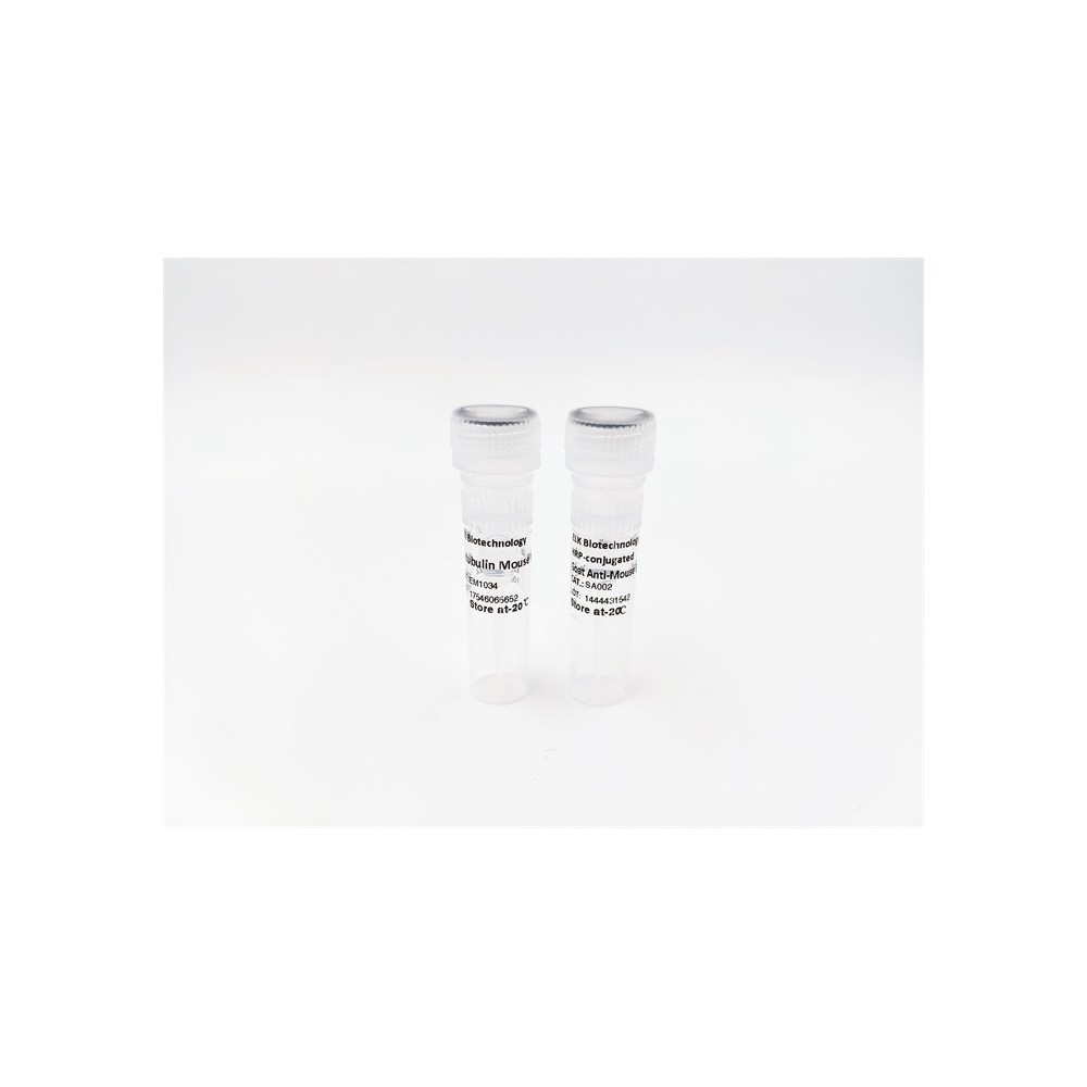

| ELK.No | ES13035 |
| Product name | SNTA1 rabbit pAb |
| Reactivity | Human; Mouse |
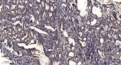
| Applications | WB;IHC |
| Other name | |
| Size | 100μL |
| Unit price ($) | 248 |
| Human gene ID | 6640 |
| Human Swiss-Prot | Q13424 |
| Source | Rabbit |
| Isotype | IgG |
| Target | SNTA1 |
| Fields | |
| Gene name | SNTA1 SNT1 |
| Protein name | SNTA1 |
| Human gene link | View Human Gene |
| Human Swiss link | View Human Swiss-Prot |
| Mouse gene ID | |
| Mouse gene link | |
| Mouse Swiss-Prot | Q61234 |
| Mouse Swiss link | View Mouse Swiss-Prot |
| Rat gene ID | |
| Rat gene link | |
| Rat Swiss-Prot | |
| Rat Swiss link | |
| Immunogen | Synthesized peptide derived from human SNTA1 AA range: 289-339 |
| Specificity | This antibody detects endogenous levels of SNTA1 at Human/Mouse |
| Formulation | Liquid in PBS containing 50% glycerol, 0.5% BSA and 0.02% sodium azide. |
| Clonality | Polyclonal |
| Dilution | WB 1:500-2000;IHC-p 1:50-300 |
| Purification | The antibody was affinity-purified from rabbit serum by affinity-chromatography using specific immunogen. |
| Concentration | 1 mg/ml |
| Storage stability | -20°C/1 year |
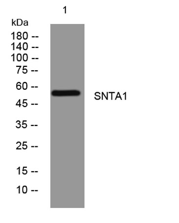
| Molecular Weight (Da) | 56kD |
| Observed band (KD) | |
| Background | Syntrophins are cytoplasmic peripheral membrane scaffold proteins that are components of the dystrophin-associated protein complex. This gene is a member of the syntrophin gene family and encodes the most common syntrophin isoform found in cardiac tissues. The N-terminal PDZ domain of this syntrophin protein interacts with the C-terminus of the pore-forming alpha subunit (SCN5A) of the cardiac sodium channel Nav1.5. This protein also associates cardiac sodium channels with the nitric oxide synthase-PMCA4b (plasma membrane Ca-ATPase subtype 4b) complex in cardiomyocytes. This gene is a susceptibility locus for Long-QT syndrome (LQT) - an inherited disorder associated with sudden cardiac death from arrhythmia - and sudden infant death syndrome (SIDS). This protein also associates with dystrophin and dystrophin-related proteins at the neuromuscular junction and alters intracellular calcium ion levels in muscle tissue. [provided by RefSeq, Jan 2013], |
| Function | domain:The PDZ domain binds to the last three or four amino acids of ion channels and receptor proteins. The association with dystrophin or related proteins probably leaves the PDZ domain available to recruit proteins to the membrane.,domain:The PH 1 domain mediates the oligomerization in a calcium dependent manner, and the association with the phosphatidylinositol 4,5-biphosphate.,domain:The SU domain binds calmodulin in a calcium-dependent manner.,function:Adapter protein that binds to and probably organizes the subcellular localization of a variety of membrane proteins. May link various receptors to the actin cytoskeleton and the extracellular matrix via the dystrophin glycoprotein complex. Plays an important role in synapse formation and in the organization of UTRN and acetylcholine receptors at the neuromuscular synapse. Binds to phosphatidylinositol 4,5-biphosphate.,PTM:Phosphoryla |
| Subcellular location | Cell membrane, sarcolemma ; Peripheral membrane protein ; Cytoplasmic side . Cell junction . Cytoplasm, cytoskeleton . In skeletal muscle, it localizes at the cytoplasmic side of the sarcolemmal membrane and at neuromuscular junctions. . |
| Expression | High expression in skeletal muscle and heart. Low expression in brain, pancreas, liver, kidney and lung. Not detected in placenta. |

Use collapsible tabs for more detailed information that will help customers make a purchasing decision.
Ex: Shipping and return policies, size guides, and other common questions.