 Français
 English

| ELK.No | ES13212 |
| Product name | SALL2 rabbit pAb |
| Reactivity | Human; Mouse |
| Applications | WB |
| Other name | |
| Size | 50μL |
| Unit price ($) | 148 |
| Human gene ID | 6297 |
| Human Swiss-Prot | Q9Y467 |
| Source | Rabbit |
| Isotype | IgG |
| Target | SALL2 |
| Fields | |
| Gene name | SALL2 KIAA0360 SAL2 ZNF795 |
| Protein name | SALL2 |
| Human gene link | View Human Gene |
| Human Swiss link | View Human Swiss-Prot |
| Mouse gene ID | 50524 |
| Mouse gene link | View Mouse Gene |
| Mouse Swiss-Prot | Q9QX96 |
| Mouse Swiss link | View Mouse Swiss-Prot |
| Rat gene ID | |
| Rat gene link | |
| Rat Swiss-Prot | |
| Rat Swiss link | |
| Immunogen | Synthesized peptide derived from human SALL2 AA range: 956-1006 |
| Specificity | This antibody detects endogenous levels of SALL2 at Human/Mouse |
| Formulation | Liquid in PBS containing 50% glycerol, 0.5% BSA and 0.02% sodium azide. |
| Clonality | Polyclonal |
| Dilution | WB 1:500-2000 |
| Purification | The antibody was affinity-purified from rabbit serum by affinity-chromatography using specific immunogen. |
| Concentration | 1 mg/ml |
| Storage stability | -20°C/1 year |
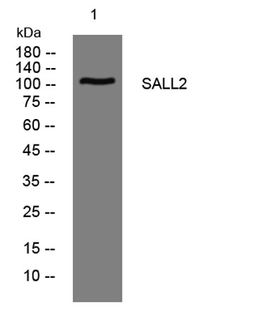
| Molecular Weight (Da) | 111kD |
| Observed band (KD) | |
| Background | This gene encodes a protein containing multiple zinc finger domains. The encoded protein functions in optical fissure closure during development of the eye in the embryo. Mutations in this gene are associated with ocular coloboma. [provided by RefSeq, Jul 2016], |
| Function | developmental stage:In fetal brain exclusively in pontine nuclei.,function:Probable transcription factor.,similarity:Belongs to the sal C2H2-type zinc-finger protein family.,similarity:Contains 7 C2H2-type zinc fingers.,tissue specificity:Highest levels in adult brain (in different areas). Lower levels in heart; very low levels in kidney and pancreas., |
| Subcellular location | Nucleus . |
| Expression | Highest levels in adult brain (in different areas). Lower levels in heart; very low levels in kidney and pancreas. Expressed throughout the retina and lens vesicle as well as the periocular mesenchyme. |

Use collapsible tabs for more detailed information that will help customers make a purchasing decision.
Ex: Shipping and return policies, size guides, and other common questions.