 Français
 English
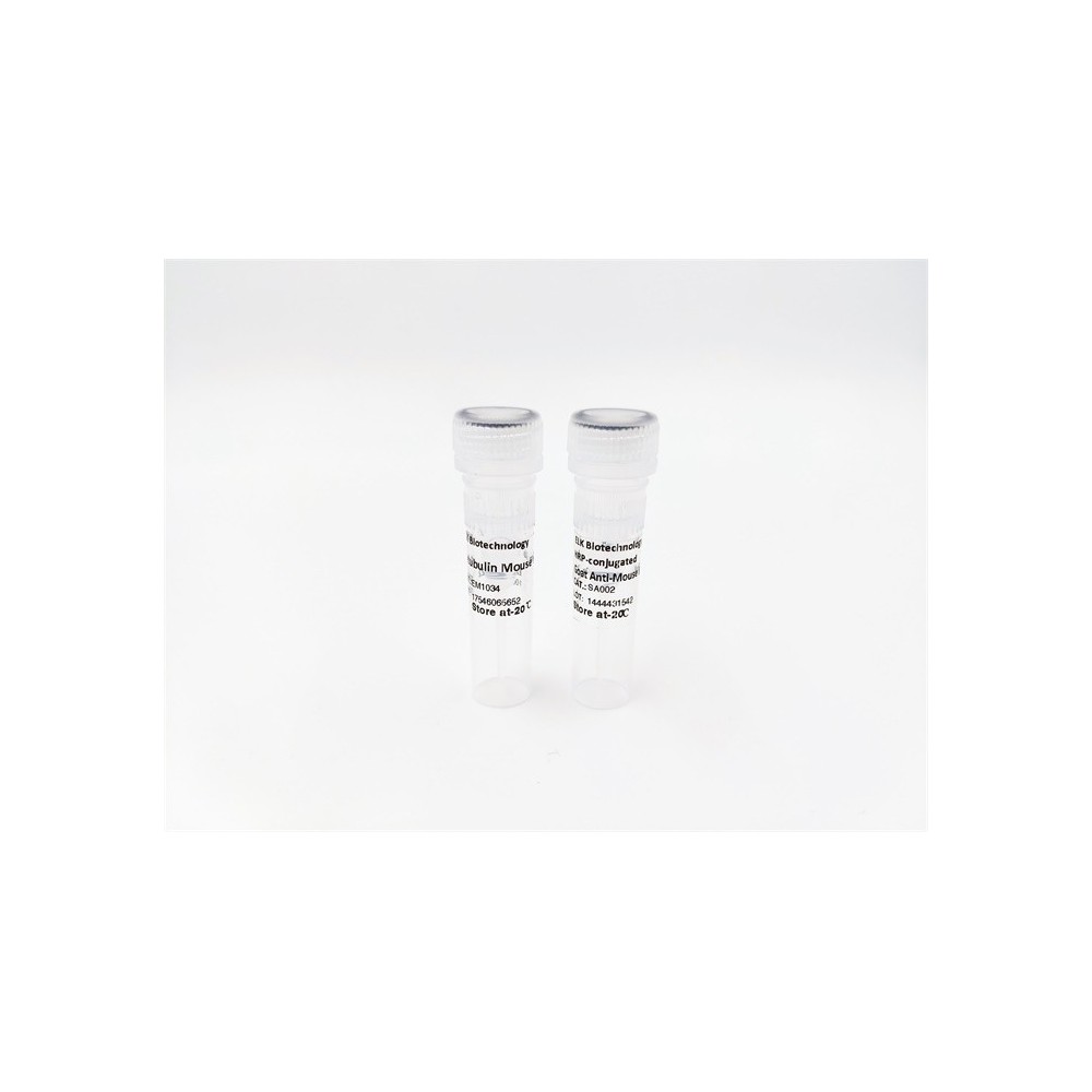

| ELK.No | ES13403 |
| Product name | RHBT2 rabbit pAb |
| Reactivity | Human; Mouse |
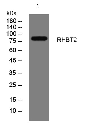
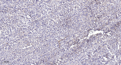
| Applications | WB;ELISA;IHC |
| Other name | |
| Size | 50μL |
| Unit price ($) | 148 |
| Human gene ID | 23221 |
| Human Swiss-Prot | Q9BYZ6 |
| Source | Rabbit |
| Isotype | IgG |
| Target | RHBT2 |
| Fields | >>Ubiquitin mediated proteolysis |
| Gene name | RHOBTB2 DBC2 KIAA0717 |
| Protein name | RHBT2 |
| Human gene link | View Human Gene |
| Human Swiss link | View Human Swiss-Prot |
| Mouse gene ID | 246710 |
| Mouse gene link | View Mouse Gene |
| Mouse Swiss-Prot | Q91V93 |
| Mouse Swiss link | View Mouse Swiss-Prot |
| Rat gene ID | |
| Rat gene link | |
| Rat Swiss-Prot | |
| Rat Swiss link | |
| Immunogen | Synthesized peptide derived from human RHBT2 AA range: 573-623 |
| Specificity | This antibody detects endogenous levels of RHBT2 at Human/Mouse |
| Formulation | Liquid in PBS containing 50% glycerol, 0.5% BSA and 0.02% sodium azide. |
| Clonality | Polyclonal |
| Dilution | WB 1:500-2000;IHC-p 1:50-300; ELISA 2000-20000 |
| Purification | The antibody was affinity-purified from rabbit serum by affinity-chromatography using specific immunogen. |
| Concentration | 1 mg/ml |
| Storage stability | -20°C/1 year |
| Molecular Weight (Da) | 80kD |
| Observed band (KD) | |
| Background | The protein encoded by this gene is a small Rho GTPase and a candidate tumor suppressor. The encoded protein interacts with the cullin-3 protein, a ubiquitin E3 ligase necessary for mitotic cell division. This protein inhibits the growth and spread of some types of breast cancer. Three transcript variants encoding different isoforms have been found for this gene. [provided by RefSeq, Nov 2011], |
| Function | similarity:Belongs to the small GTPase superfamily. Rho family.,similarity:Contains 2 BTB (POZ) domains.,tissue specificity:Ubiquitous, with highest levels in neural tissues. Expression is also detected in fetal lung, heart, and brain., |
| Subcellular location | intracellular,cytosol,plasma membrane, |
| Expression | Ubiquitous, with highest levels in neural tissues. Expression is also detected in fetal lung, heart, and brain. |

Use collapsible tabs for more detailed information that will help customers make a purchasing decision.
Ex: Shipping and return policies, size guides, and other common questions.