 Français
 English

| ELK.No | ES13432 |
| Product name | RD3 rabbit pAb |
| Reactivity | Human; Mouse |
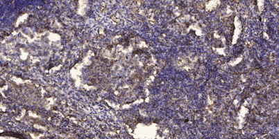
| Applications | WB;ELISA;IHC |
| Other name | |
| Size | 50μL |
| Unit price ($) | 148 |
| Human gene ID | 343035 |
| Human Swiss-Prot | Q7Z3Z2 |
| Source | Rabbit |
| Isotype | IgG |
| Target | RD3 |
| Fields | |
| Gene name | RD3 C1orf36 |
| Protein name | RD3 |
| Human gene link | View Human Gene |
| Human Swiss link | View Human Swiss-Prot |
| Mouse gene ID | 74023 |
| Mouse gene link | View Mouse Gene |
| Mouse Swiss-Prot | Q8BRE0 |
| Mouse Swiss link | View Mouse Swiss-Prot |
| Rat gene ID | |
| Rat gene link | |
| Rat Swiss-Prot | |
| Rat Swiss link | |
| Immunogen | Synthesized peptide derived from human RD3 AA range: 126-176 |
| Specificity | This antibody detects endogenous levels of RD3 at Human/Mouse |
| Formulation | Liquid in PBS containing 50% glycerol, 0.5% BSA and 0.02% sodium azide. |
| Clonality | Polyclonal |
| Dilution | WB 1:500-2000;IHC-p 1:50-300; ELISA 2000-20000 |
| Purification | The antibody was affinity-purified from rabbit serum by affinity-chromatography using specific immunogen. |
| Concentration | 1 mg/ml |
| Storage stability | -20°C/1 year |
| Molecular Weight (Da) | 21kD |
| Observed band (KD) | |
| Background | This gene encodes a retinal protein that is associated with promyelocytic leukemia-gene product (PML) bodies in the nucleus. Mutations in this gene cause Leber congenital amaurosis type 12, a disease that results in retinal degeneration. Alternative splicing results in multiple transcript variants. [provided by RefSeq, Sep 2009], |
| Function | disease:Defects in RD3 are the cause of Leber congenital amaurosis type 12 (LCA12) [MIM:610612]. LCA designates a clinically and genetically heterogeneous group of childhood retinal degenerations, generally inherited in an autosomal recessive manner. Affected infants have little or no retinal photoreceptor function as tested by electroretinography. LCA represents the most common genetic cause of congenital visual impairment in infants and children.,tissue specificity:Preferentially expressed in retina., |
| Subcellular location | Cell projection, cilium, photoreceptor outer segment . Photoreceptor inner segment . Endosome . Nucleus . Cytoplasm . Cytoplasm, perinuclear region . Colocalizes with GUCY2E and GUCY2F in rods and cones photoreceptors. Colocalizes with GUK1 in photoreceptor inner segments and to a lesser extent in the outer plexiform layer (By similarity). Strong dot-like perinuclear staining in the epithelial cells (PubMed:29030614). . |
| Expression | Expressed in retina (PubMed:12914764). Widely expressed (at protein level) (PubMed:29030614). In the retina the strongest immunoreactivity is detected in the inner half of the cytoplasmic portion of the photoreceptor layer, where rods and cones are found, and the external half of the outer plexiform layer (at protein level) (PubMed:29030614). |


Use collapsible tabs for more detailed information that will help customers make a purchasing decision.
Ex: Shipping and return policies, size guides, and other common questions.