 Français
 English
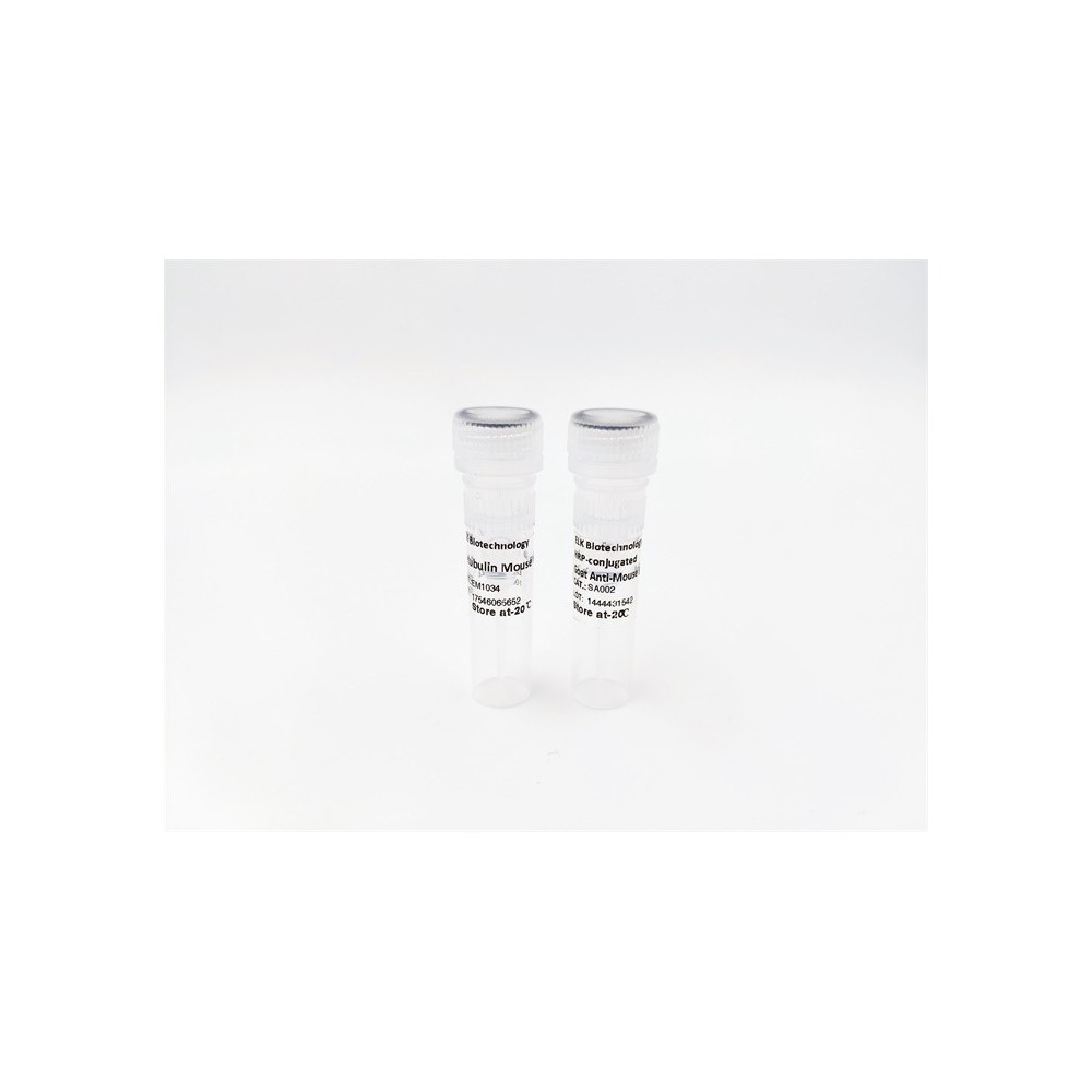
RCC1 (phospho-Ser11) rabbit pAb
AO-06-ES13434-100

| ELK.No | ES13434 |
| Product name | RCC1 (phospho-Ser11) rabbit pAb |
| Reactivity | Human;Rat;Mouse; |
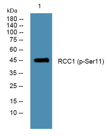
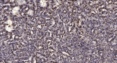
| Applications | WB;IHC |
| Other name | Regulator of chromosome condensation (Cell cycle regulatory protein) (Chromosome condensation protein 1) |
| Size | 100μL |
| Unit price ($) | 248 |
| Human gene ID | 1104 |
| Human Swiss-Prot | P18754 |
| Source | Rabbit |
| Isotype | IgG |
| Target | RCC1 |
| Fields | |
| Gene name | RCC1 CHC1 |
| Protein name | RCC1 (Ser11) |
| Human gene link | View Human Gene |
| Human Swiss link | View Human Swiss-Prot |
| Mouse gene ID | 100088 |
| Mouse gene link | View Mouse Gene |
| Mouse Swiss-Prot | Q8VE37 |
| Mouse Swiss link | View Mouse Swiss-Prot |
| Rat gene ID | |
| Rat gene link | |
| Rat Swiss-Prot | |
| Rat Swiss link | |
| Immunogen | Synthesized phosho peptide around human RCC1 (Ser11) |
| Specificity | This antibody detects endogenous levels of Human RCC1 (phospho-Ser11) |
| Formulation | Liquid in PBS containing 50% glycerol, 0.5% BSA and 0.02% sodium azide. |
| Clonality | Polyclonal |
| Dilution | WB 1:500-2000;IHC-p 1:50-300 |
| Purification | The antibody was affinity-purified from rabbit serum by affinity-chromatography using specific immunogen. |
| Concentration | 1 mg/ml |
| Storage stability | -20°C/1 year |
| Molecular Weight (Da) | |
| Observed band (KD) | 45kD |
| Background | |
| Function | disease:Patients with Raynaud disease produce antibodies that bind to RCC1.,function:Promotes the exchange of Ran-bound GDP by GTP. Involved in the regulation of onset of chromosome condensation in the S phase. Binds to the chromatin. RCC1/Ran complex (together with other proteins) acts as a component of a signal transmission pathway that detects unreplicated DNA.,similarity:Contains 7 RCC1 repeats.,subcellular location:Becomes dispersed throughout the cytoplasm during mitosis., |
| Subcellular location | Nucleus . Chromosome . Cytoplasm . Predominantly nuclear in interphase cells (PubMed:12194828). Binds to mitotic chromosomes (PubMed:12194828, PubMed:17435751, PubMed:20668449). . |
| Expression | Brain,Breast,Epithelium,Eye,Muscle,Testis, |

Use collapsible tabs for more detailed information that will help customers make a purchasing decision.
Ex: Shipping and return policies, size guides, and other common questions.