 Français
 English

| ELK.No | ES13437 |
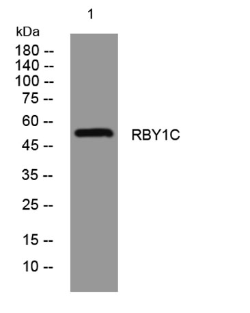
| Product name | RBY1C rabbit pAb |
| Reactivity | Human;Rat;Mouse; |
| Applications | WB |
| Other name | |
| Size | 50μL |
| Unit price ($) | 148 |
| Human gene ID | |
| Human Swiss-Prot | P0DJD4 |
| Source | Rabbit |
| Isotype | IgG |
| Target | RBY1C |
| Fields | |
| Gene name | RBMY1C |
| Protein name | RBY1C |
| Human gene link | |
| Human Swiss link | View Human Swiss-Prot |
| Mouse gene ID | |
| Mouse gene link | |
| Mouse Swiss-Prot | |
| Mouse Swiss link | |
| Rat gene ID | |
| Rat gene link | |
| Rat Swiss-Prot | |
| Rat Swiss link | |
| Immunogen | Synthesized peptide derived from human RBY1C AA range: 321-371 |
| Specificity | This antibody detects endogenous levels of RBY1C at Human |
| Formulation | Liquid in PBS containing 50% glycerol, 0.5% BSA and 0.02% sodium azide. |
| Clonality | Polyclonal |
| Dilution | WB 1:500-2000 |
| Purification | The antibody was affinity-purified from rabbit serum by affinity-chromatography using specific immunogen. |
| Concentration | 1 mg/ml |
| Storage stability | -20°C/1 year |
| Molecular Weight (Da) | 55kD |
| Observed band (KD) | |
| Background | |
| Function | developmental stage:Expressed in all of the transcriptionally active stages of germ cell development from spermatogonia through spermatocytes to round spermatids.,function:RNA-binding protein which may be involved in spermatogenesis. Required for sperm development, possibly by participating in pre-mRNA splicing in the testis.,miscellaneous:The RBMY1 proteins are encoded by strongly repeated regions of the Y chromosome, mostly within the AZFb region. The exact number of functional copies is unclear and may vary between individues, and some of them may represent pseudogenes. The proteins are very similar, which gives their indidual characterization difficult. Thus, most experiments do not discriminate between the different members. One can therefore suppose that reported interactions with a RBMY1 protein involve all the proteins.,similarity:Contains 1 RRM (RNA recognition motif) domain.,su |
| Subcellular location | Nucleus . |
| Expression | Testis-specific. |

Use collapsible tabs for more detailed information that will help customers make a purchasing decision.
Ex: Shipping and return policies, size guides, and other common questions.