 Français
 English
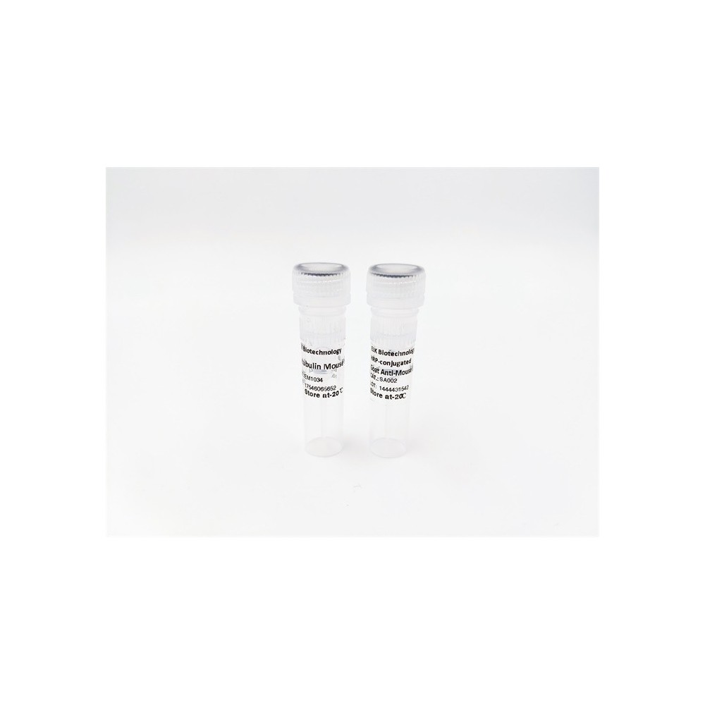

| ELK.No | ES13443 |
| Product name | RBMS2 rabbit pAb |
| Reactivity | Human; Mouse |
| Applications | WB;IHC |
| Other name | |
| Size | 100μL |
| Unit price ($) | 248 |
| Human gene ID | 5939 |
| Human Swiss-Prot | Q15434 |
| Source | Rabbit |
| Isotype | IgG |
| Target | RBMS2 |
| Fields | |
| Gene name | RBMS2 SCR3 |
| Protein name | RBMS2 |
| Human gene link | View Human Gene |
| Human Swiss link | View Human Swiss-Prot |
| Mouse gene ID | 56516 |
| Mouse gene link | View Mouse Gene |
| Mouse Swiss-Prot | Q8VC70 |
| Mouse Swiss link | View Mouse Swiss-Prot |
| Rat gene ID | |
| Rat gene link | |
| Rat Swiss-Prot | |
| Rat Swiss link | |
| Immunogen | Synthesized peptide derived from human RBMS2 AA range: 194-244 |
| Specificity | This antibody detects endogenous levels of RBMS2 at Human/Mouse |
| Formulation | Liquid in PBS containing 50% glycerol, 0.5% BSA and 0.02% sodium azide. |
| Clonality | Polyclonal |
| Dilution | WB 1:500-2000;IHC-p 1:50-300 |
| Purification | The antibody was affinity-purified from rabbit serum by affinity-chromatography using specific immunogen. |
| Concentration | 1 mg/ml |
| Storage stability | -20°C/1 year |
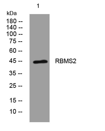
| Molecular Weight (Da) | 45kD |
| Observed band (KD) | |
| Background | The protein encoded by this gene is a member of a small family of proteins which bind single stranded DNA/RNA. These proteins are characterized by the presence of two sets of ribonucleoprotein consensus sequence (RNP-CS) that contain conserved motifs, RNP1 and RNP2, originally described in RNA binding proteins, and required for DNA binding. The RBMS proteins have been implicated in such diverse functions as DNA replication, gene transcription, cell cycle progression and apoptosis. This protein was isolated by phenotypic complementation of cdc2 and cdc13 mutants of yeast and is thought to suppress cdc2 and cdc13 mutants through the induction of translation of cdc2. [provided by RefSeq, Jul 2008], |
| Function | similarity:Contains 2 RRM (RNA recognition motif) domains., |
| Subcellular location | Nucleus . |
| Expression |

Use collapsible tabs for more detailed information that will help customers make a purchasing decision.
Ex: Shipping and return policies, size guides, and other common questions.