 Français
 English
NFκB-p65 (phospho Ser529) rabbit pAb
AO-06-ES1370-100

| ELK.No | ES1370 |
| Product name | NFκB-p65 (phospho Ser529) rabbit pAb |
| Reactivity | Human;Mouse;Rat;Monkey |
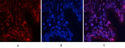
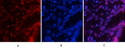
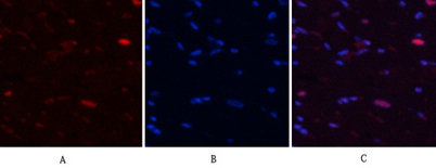
| Applications | IF;WB;IHC;ELISA |
| Other name | RELA; NFKB3; Transcription factor p65; Nuclear factor NF-kappa-B p65 subunit; Nuclear factor of kappa light polypeptide gene enhancer in B-cells 3 |
| Size | 100μL |
| Unit price ($) | 248 |
| Human gene ID | 5970 |
| Human Swiss-Prot | Q04206 |
| Source | Rabbit |
| Isotype | IgG |
| Target | NFkB p65 |
| Fields | >>Antifolate resistance;>>MAPK signaling pathway;>>Ras signaling pathway;>>cAMP signaling pathway;>>Chemokine signaling pathway;>>NF-kappa B signaling pathway;>>HIF-1 signaling pathway;>>Sphingolipid signaling pathway;>>Mitophagy - animal;>>PI3K-Akt signaling pathway;>>Apoptosis;>>Longevity regulating pathway;>>Cellular senescence;>>Osteoclast differentiation;>>Neutrophil extracellular trap formation;>>Toll-like receptor signaling pathway;>>NOD-like receptor signaling pathway;>>RIG-I-like receptor signaling pathway;>>Cytosolic DNA-sensing pathway;>>C-type lectin receptor signaling pathway;>>IL-17 signaling pathway;>>Th1 and Th2 cell differentiation;>>Th17 cell differentiation;>>T cell receptor signaling pathway;>>B cell receptor signaling pathway;>>TNF signaling pathway;>>Neurotrophin signaling pathway;>>Prolactin signaling pathway;>>Adipocytokine signaling pathway;>>Relaxin signaling pathway;>>Insulin resistance;>>Non-alcoholic fatty liver disease;>>AGE-RAGE signaling pathway in diabe |
| Gene name | RELA |
| Protein name | Transcription factor p65 |
| Human gene link | View Human Gene |
| Human Swiss link | View Human Swiss-Prot |
| Mouse gene ID | 19697 |
| Mouse gene link | View Mouse Gene |
| Mouse Swiss-Prot | Q04207 |
| Mouse Swiss link | View Mouse Swiss-Prot |
| Rat gene ID | |
| Rat gene link | |
| Rat Swiss-Prot | |
| Rat Swiss link | |
| Immunogen | Synthesized phospho-peptide around the phosphorylation site of human NFκB-p65 (phospho Ser529) |
| Specificity | Phospho-NFκB-p65 (S529) Polyclonal Antibody detects endogenous levels of NFκB-p65 protein only when phosphorylated at S529. |
| Formulation | Liquid in PBS containing 50% glycerol, 0.5% BSA and 0.02% sodium azide. |
| Clonality | Polyclonal |
| Dilution | IF: 1:50-200 Western Blot: 1/500 - 1/2000. Immunohistochemistry: 1/100 - 1/300. ELISA: 1/20000. Not yet tested in other applications. |
| Purification | The antibody was affinity-purified from rabbit antiserum by affinity-chromatography using epitope-specific immunogen. |
| Concentration | 1 mg/ml |
| Storage stability | -20°C/1 year |
| Molecular Weight (Da) | |
| Observed band (KD) | 60kD |
| Background | NF-kappa-B is a ubiquitous transcription factor involved in several biological processes. It is held in the cytoplasm in an inactive state by specific inhibitors. Upon degradation of the inhibitor, NF-kappa-B moves to the nucleus and activates transcription of specific genes. NF-kappa-B is composed of NFKB1 or NFKB2 bound to either REL, RELA, or RELB. The most abundant form of NF-kappa-B is NFKB1 complexed with the product of this gene, RELA. Four transcript variants encoding different isoforms have been found for this gene. [provided by RefSeq, Sep 2011], |
| Function | function:NF-kappa-B is a pleiotropic transcription factor which is present in almost all cell types and is involved in many biological processed such as inflammation, immunity, differentiation, cell growth, tumorigenesis and apoptosis. NF-kappa-B is a homo- or heterodimeric complex formed by the Rel-like domain-containing proteins RELA/p65, RELB, NFKB1/p105, NFKB1/p50, REL and NFKB2/p52 and the heterodimeric p65-p50 complex appears to be most abundant one. The dimers bind at kappa-B sites in the DNA of their target genes and the individual dimers have distinct preferences for different kappa-B sites that they can bind with distinguishable affinity and specificity. Different dimer combinations act as transcriptional activators or repressors, respectively. NF-kappa-B is controlled by various mechanisms of post-translational modification and subcellular compartmentalization as well as by in |
| Subcellular location | Nucleus . Cytoplasm . Nuclear, but also found in the cytoplasm in an inactive form complexed to an inhibitor (I-kappa-B) (PubMed:1493333). Colocalized with DDX1 in the nucleus upon TNF-alpha induction (PubMed:19058135). Colocalizes with GFI1 in the nucleus after LPS stimulation (PubMed:20547752). Translocation to the nucleus is impaired in L.monocytogenes infection (PubMed:20855622). . |
| Expression | Bone,Colon,Pancreas,Placenta, |

Use collapsible tabs for more detailed information that will help customers make a purchasing decision.
Ex: Shipping and return policies, size guides, and other common questions.