 Français
 English
p53 (phospho Thr18) rabbit pAb
AO-06-ES1388-100

| ELK.No | ES1388 |
| Product name | p53 (phospho Thr18) rabbit pAb |
| Reactivity | Human;Mouse;Rat |
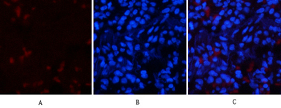
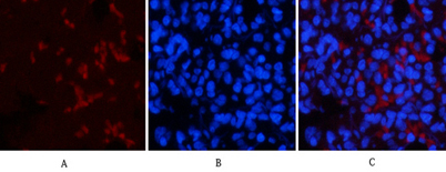
| Applications | IF;WB;IHC;ELISA |
| Other name | TP53; P53; Cellular tumor antigen p53; Antigen NY-CO-13; Phosphoprotein p53; Tumor suppressor p53 |
| Size | 100μL |
| Unit price ($) | 248 |
| Human gene ID | 7157 |
| Human Swiss-Prot | P04637 |
| Source | Rabbit |
| Isotype | IgG |
| Target | p53 |
| Fields | >>Endocrine resistance;>>Platinum drug resistance;>>MAPK signaling pathway;>>Sphingolipid signaling pathway;>>Cell cycle;>>p53 signaling pathway;>>Mitophagy - animal;>>PI3K-Akt signaling pathway;>>Apoptosis;>>Longevity regulating pathway;>>Ferroptosis;>>Cellular senescence;>>Wnt signaling pathway;>>Neurotrophin signaling pathway;>>Thyroid hormone signaling pathway;>>Parkinson disease;>>Amyotrophic lateral sclerosis;>>Huntington disease;>>Shigellosis;>>Hepatitis C;>>Hepatitis B;>>Measles;>>Human cytomegalovirus infection;>>Human papillomavirus infection;>>Human T-cell leukemia virus 1 infection;>>Kaposi sarcoma-associated herpesvirus infection;>>Herpes simplex virus 1 infection;>>Epstein-Barr virus infection;>>Pathways in cancer;>>Transcriptional misregulation in cancer;>>Viral carcinogenesis;>>Proteoglycans in cancer;>>MicroRNAs in cancer;>>Colorectal cancer;>>Pancreatic cancer;>>Endometrial cancer;>>Glioma;>>Prostate cancer;>>Thyroid cancer;>>Basal cell carcinoma;>>Melanoma;>>Bladder |
| Gene name | TP53 |
| Protein name | Cellular tumor antigen p53 |
| Human gene link | View Human Gene |
| Human Swiss link | View Human Swiss-Prot |
| Mouse gene ID | 22059 |
| Mouse gene link | View Mouse Gene |
| Mouse Swiss-Prot | P02340 |
| Mouse Swiss link | View Mouse Swiss-Prot |
| Rat gene ID | 24842 |
| Rat gene link | View Rat Gene |
| Rat Swiss-Prot | P10361 |
| Rat Swiss link | View Rat Swiss-Prot |
| Immunogen | The antiserum was produced against synthesized peptide derived from human p53 around the phosphorylation site of Thr18. AA range:1-50 |
| Specificity | Phospho-p53 (T18) Polyclonal Antibody detects endogenous levels of p53 protein only when phosphorylated at T18. |
| Formulation | Liquid in PBS containing 50% glycerol, 0.5% BSA and 0.02% sodium azide. |
| Clonality | Polyclonal |
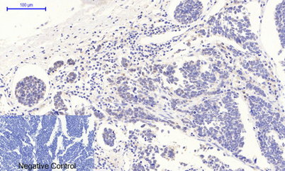
| Dilution | IF: 1:50-200 WB 1:500-2000, IHC 1:50-300 IHC 1:50-300 |
| Purification | The antibody was affinity-purified from rabbit antiserum by affinity-chromatography using epitope-specific immunogen. |
| Concentration | 1 mg/ml |
| Storage stability | -20°C/1 year |
| Molecular Weight (Da) | |
| Observed band (KD) | 46kD |
| Background | tumor protein p53(TP53) Homo sapiens This gene encodes a tumor suppressor protein containing transcriptional activation, DNA binding, and oligomerization domains. The encoded protein responds to diverse cellular stresses to regulate expression of target genes, thereby inducing cell cycle arrest, apoptosis, senescence, DNA repair, or changes in metabolism. Mutations in this gene are associated with a variety of human cancers, including hereditary cancers such as Li-Fraumeni syndrome. Alternative splicing of this gene and the use of alternate promoters result in multiple transcript variants and isoforms. Additional isoforms have also been shown to result from the use of alternate translation initiation codons (PMIDs: 12032546, 20937277). [provided by RefSeq, Feb 2013], |
| Function | cofactor:Binds 1 zinc ion per subunit.,disease:Defects in TP53 are a cause of choroid plexus papilloma [MIM:260500]. Choroid plexus papilloma is a slow-growing benign tumor of the choroid plexus that often invades the leptomeninges. In children it is usually in a lateral ventricle but in adults it is more often in the fourth ventricle. Hydrocephalus is common, either from obstruction or from tumor secretion of cerebrospinal fluid. If it undergoes malignant transformation it is called a choroid plexus carcinoma. Primary choroid plexus tumors are rare and usually occur in early childhood.,disease:Defects in TP53 are a cause of Li-Fraumeni syndrome (LFS) [MIM:151623]. LFS is an autosomal dominant familial cancer syndrome that in its classic form is defined by the existence of a proband affected by a sarcoma before 45 years with a first degree relative affected by any tumor before 45 years a |
| Subcellular location | Cytoplasm . Nucleus . Nucleus, PML body . Endoplasmic reticulum . Mitochondrion matrix . Cytoplasm, cytoskeleton, microtubule organizing center, centrosome . Recruited into PML bodies together with CHEK2 (PubMed:12810724). Translocates to mitochondria upon oxidative stress (PubMed:22726440). Translocates to mitochondria in response to mitomycin C treatment (PubMed:27323408). .; [Isoform 1]: Nucleus . Cytoplasm. Predominantly nuclear but localizes to the cytoplasm when expressed with isoform 4.; [Isoform 2]: Nucleus. Cytoplasm. Localized mainly in the nucleus with minor staining in the cytoplasm.; [Isoform 3]: Nucleus. Cytoplasm. Localized in the nucleus in most cells but found in the cytoplasm in some cells.; [Isoform 4]: Nucleus. Cytoplasm. Predominantly nuclear but translocates to the cy |
| Expression | Ubiquitous. Isoforms are expressed in a wide range of normal tissues but in a tissue-dependent manner. Isoform 2 is expressed in most normal tissues but is not detected in brain, lung, prostate, muscle, fetal brain, spinal cord and fetal liver. Isoform 3 is expressed in most normal tissues but is not detected in lung, spleen, testis, fetal brain, spinal cord and fetal liver. Isoform 7 is expressed in most normal tissues but is not detected in prostate, uterus, skeletal muscle and breast. Isoform 8 is detected only in colon, bone marrow, testis, fetal brain and intestine. Isoform 9 is expressed in most normal tissues but is not detected in brain, heart, lung, fetal liver, salivary gland, breast or intestine. |

Use collapsible tabs for more detailed information that will help customers make a purchasing decision.
Ex: Shipping and return policies, size guides, and other common questions.