 Français
 English

| ELK.No | ES13900 |
| Product name | PRS55 rabbit pAb |
| Reactivity | Human; Mouse |
| Applications | WB |
| Other name | Serine protease 55 (EC 3.4.21.-) |
| Size | 50μL |
| Unit price ($) | 148 |
| Human gene ID | 203074 |
| Human Swiss-Prot | Q6UWB4 |
| Source | Rabbit |
| Isotype | IgG |
| Target | PRS55 |
| Fields | |
| Gene name | PRSS55 UNQ9391/PRO34284 |
| Protein name | PRS55 |
| Human gene link | View Human Gene |
| Human Swiss link | View Human Swiss-Prot |
| Mouse gene ID | |
| Mouse gene link | |
| Mouse Swiss-Prot | Q14BX2 |
| Mouse Swiss link | View Mouse Swiss-Prot |
| Rat gene ID | |
| Rat gene link | |
| Rat Swiss-Prot | |
| Rat Swiss link | |
| Immunogen | Synthesized peptide derived from human PRS55 AA range: 173-223 |
| Specificity | This antibody detects endogenous levels of PRS55 at Human/Mouse |
| Formulation | Liquid in PBS containing 50% glycerol, 0.5% BSA and 0.24% sodium azide. |
| Clonality | Polyclonal |
| Dilution | WB 1:500-2000 |
| Purification | The antibody was affinity-purified from rabbit serum by affinity-chromatography using specific immunogen. |
| Concentration | 1 mg/ml |
| Storage stability | -20°C/1 year |
| Molecular Weight (Da) | |
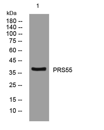
| Observed band (KD) | 38kD |
| Background | This gene encodes a member of a group of membrane-anchored chymotrypsin (S1)-like serine proteases. The enocoded protein is primarily expressed in the Leydig and Sertoli cells of the testis and may be involved in male fertility. Alternate splicing results in multiple transcript variants. [provided by RefSeq, Sep 2010], |
| Function | function:Probable serine protease.,similarity:Belongs to the peptidase S1 family.,similarity:Contains 1 peptidase S1 domain., |
| Subcellular location | [Isoform 1]: Cell membrane ; Lipid-anchor, GPI-anchor . Cytoplasm, cytosol . Mainly found in the membrane part of the cells and only in small amounts in the cytosol. .; [Isoform 2]: Cytoplasm, cytosol . Present primarily in the cytosol and only in minor amounts in the membrane fraction. . |
| Expression | Only detected in testis. Expressed in spermatogonia, spermatocytes, spermatids, Leydig and Sertoli cells. Expressed in prostate cancer and ovarian cancer (at protein level). |

Use collapsible tabs for more detailed information that will help customers make a purchasing decision.
Ex: Shipping and return policies, size guides, and other common questions.