 Français
 English
PPAR-γ (phospho Ser112) rabbit pAb
AO-06-ES1428-100

| ELK.No | ES1428 |
| Product name | PPAR-γ (phospho Ser112) rabbit pAb |
| Reactivity | Human;Mouse;Rat |
| Applications | WB;ELISA |
| Other name | PPARG; NR1C3; Peroxisome proliferator-activated receptor gamma; PPAR-gamma; Nuclear receptor subfamily 1 group C member 3 |
| Size | 100μL |
| Unit price ($) | 248 |
| Human gene ID | 5468 |
| Human Swiss-Prot | P37231 |
| Source | Rabbit |
| Isotype | IgG |
| Target | PPAR-γ |
| Fields | >>PPAR signaling pathway;>>AMPK signaling pathway;>>Longevity regulating pathway;>>Osteoclast differentiation;>>Thermogenesis;>>Non-alcoholic fatty liver disease;>>Huntington disease;>>Pathways in cancer;>>Transcriptional misregulation in cancer;>>Thyroid cancer;>>Lipid and atherosclerosis |
| Gene name | PPARG |
| Protein name | Peroxisome proliferator-activated receptor gamma |
| Human gene link | View Human Gene |
| Human Swiss link | View Human Swiss-Prot |
| Mouse gene ID | 19016 |
| Mouse gene link | View Mouse Gene |
| Mouse Swiss-Prot | P37238 |
| Mouse Swiss link | View Mouse Swiss-Prot |
| Rat gene ID | 25664 |
| Rat gene link | View Rat Gene |
| Rat Swiss-Prot | O88275 |
| Rat Swiss link | View Rat Swiss-Prot |
| Immunogen | The antiserum was produced against synthesized peptide derived from human PPAR-gamma around the phosphorylation site of Ser112. AA range:78-127 |
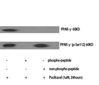
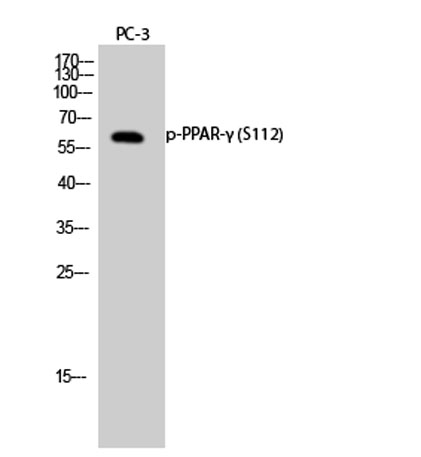
| Specificity | Phospho-PPAR-γ (S112) Polyclonal Antibody detects endogenous levels of PPAR-γ protein only when phosphorylated at S112. |
| Formulation | Liquid in PBS containing 50% glycerol, 0.5% BSA and 0.02% sodium azide. |
| Clonality | Polyclonal |
| Dilution | Western Blot: 1/500 - 1/2000. ELISA: 1/10000. Not yet tested in other applications. |
| Purification | The antibody was affinity-purified from rabbit antiserum by affinity-chromatography using epitope-specific immunogen. |
| Concentration | 1 mg/ml |
| Storage stability | -20°C/1 year |
| Molecular Weight (Da) | |
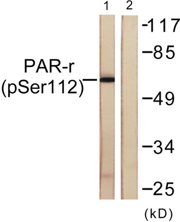
| Observed band (KD) | 60kD |
| Background | peroxisome proliferator activated receptor gamma(PPARG) Homo sapiens This gene encodes a member of the peroxisome proliferator-activated receptor (PPAR) subfamily of nuclear receptors. PPARs form heterodimers with retinoid X receptors (RXRs) and these heterodimers regulate transcription of various genes. Three subtypes of PPARs are known: PPAR-alpha, PPAR-delta, and PPAR-gamma. The protein encoded by this gene is PPAR-gamma and is a regulator of adipocyte differentiation. Additionally, PPAR-gamma has been implicated in the pathology of numerous diseases including obesity, diabetes, atherosclerosis and cancer. Alternatively spliced transcript variants that encode different isoforms have been described. [provided by RefSeq, Jul 2008], |
| Function | alternative products:Additional isoforms seem to exist,disease:Defects in PPARG are the cause of familial partial lipodystrophy type 3 (FPLD3) [MIM:604367]. Familial partial lipodystrophies (FPLD) are a heterogeneous group of genetic disorders characterized by marked loss of subcutaneous (sc) fat from the extremities. Affected individuals show an increased preponderance of insulin resistance, diabetes mellitus and dyslipidemia.,disease:Defects in PPARG can lead to type 2 insulin-resistant diabetes and hyptertension.,disease:Defects in PPARG may be associated with colon cancer.,disease:Defects in PPARG may be associated with susceptibility to obesity [MIM:601665].,disease:Variation in PPARG is associated with carotid intimal medial thickness 1 (CIMT1) [MIM:609338]. CIMT is a measure of atherosclerosis that is independently associated with traditional atherosclerotic cardiovascular disease |
| Subcellular location | Nucleus. Cytoplasm. Redistributed from the nucleus to the cytosol through a MAP2K1/MEK1-dependent manner. NOCT enhances its nuclear translocation. |
| Expression | Highest expression in adipose tissue. Lower in skeletal muscle, spleen, heart and liver. Also detectable in placenta, lung and ovary. |

Use collapsible tabs for more detailed information that will help customers make a purchasing decision.
Ex: Shipping and return policies, size guides, and other common questions.