 Français
 English

| ELK.No | ES14367 |
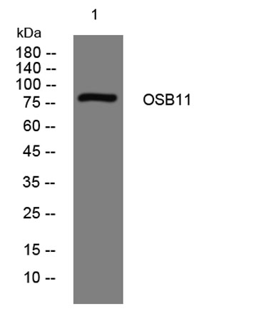
| Product name | OSB11 rabbit pAb |
| Reactivity | Human; Mouse |
| Applications | WB |
| Other name | |
| Size | 50μL |
| Unit price ($) | 148 |
| Human gene ID | 114885 |
| Human Swiss-Prot | Q9BXB4 |
| Source | Rabbit |
| Isotype | IgG |
| Target | OSB11 |
| Fields | |
| Gene name | OSBPL11 ORP11 OSBP12 |
| Protein name | OSB11 |
| Human gene link | View Human Gene |
| Human Swiss link | View Human Swiss-Prot |
| Mouse gene ID | |
| Mouse gene link | |
| Mouse Swiss-Prot | Q8CI95 |
| Mouse Swiss link | View Mouse Swiss-Prot |
| Rat gene ID | |
| Rat gene link | |
| Rat Swiss-Prot | |
| Rat Swiss link | |
| Immunogen | Synthesized peptide derived from human OSB11 AA range: 180-230 |
| Specificity | This antibody detects endogenous levels of OSB11 at Human/Mouse |
| Formulation | Liquid in PBS containing 50% glycerol, 0.5% BSA and 0.02% sodium azide. |
| Clonality | Polyclonal |
| Dilution | WB 1:500-2000 |
| Purification | The antibody was affinity-purified from rabbit serum by affinity-chromatography using specific immunogen. |
| Concentration | 1 mg/ml |
| Storage stability | -20°C/1 year |
| Molecular Weight (Da) | 82kD |
| Observed band (KD) | |
| Background | This gene encodes a member of the oxysterol-binding protein (OSBP) family, a group of intracellular lipid receptors. Like most members, the encoded protein contains an N-terminal pleckstrin homology domain and a highly conserved C-terminal OSBP-like sterol-binding domain. [provided by RefSeq, Jul 2008], |
| Function | similarity:Belongs to the OSBP family.,similarity:Contains 1 PH domain.,tissue specificity:Widely expressed., |
| Subcellular location | Late endosome membrane . Golgi apparatus, trans-Golgi network membrane . Localizes at the Golgi-late endosome interface. |
| Expression | Present at highest levels in ovary, testis, kidney, liver, stomach, brain, and adipose tissue. Strong expression (at protein level) in epithelial cells of kidney tubules, testicular tubules, caecum, and skin (PubMed:20599956). Present at low levels in subcutaneous and visceral adipose tissue (at protein level)(PubMed:23028956). |

Use collapsible tabs for more detailed information that will help customers make a purchasing decision.
Ex: Shipping and return policies, size guides, and other common questions.