 Français
 English

| ELK.No | ES13280 |
| Product name | S100A7 Rabbit pAb |
| Reactivity | Human;Rat;Mouse; |
| Applications | WB;IHC;IF;ELISA |
| Other name | Protein S100-A7 (Psoriasin;S100 calcium-binding protein A7) |
| Size | 50μL |
| Unit price ($) | 148 |
| Human gene ID | 6278 |
| Human Swiss-Prot | P31151 |
| Source | Rabbit |
| Isotype | IgG |
| Target | S100A7 |
| Fields | >>IL-17 signaling pathway |
| Gene name | S100A7 PSOR1 S100A7C |
| Protein name | S100A7 |
| Human gene link | |
| Human Swiss link | View Human Swiss-Prot |
| Mouse gene ID | |
| Mouse gene link | |
| Mouse Swiss-Prot | |
| Mouse Swiss link | |
| Rat gene ID | |
| Rat gene link | |
| Rat Swiss-Prot | |
| Rat Swiss link | |
| Immunogen | Synthesized peptide derived from human S100A7 AA range: 1-51 |
| Specificity | This antibody detects endogenous levels of human S100A7 |
| Formulation | Liquid in PBS containing 50% glycerol, 0.5% BSA and 0.02% sodium azide. |
| Clonality | Polyclonal |
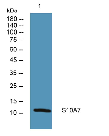
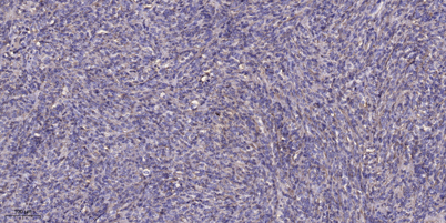
| Dilution | Western Blot: 1/500 - 1/2000. Immunohistochemistry: 1/100 - 1/300. Immunofluorescence: 1/200 - 1/1000. ELISA: 1/5000. Not yet tested in other applications. |
| Purification | The antibody was affinity-purified from mouse ascites by affinity-chromatography using specific immunogen. |
| Concentration | 1 mg/ml |
| Storage stability | -20°C/1 year |
| Molecular Weight (Da) | |
| Observed band (KD) | |
| Background | S100 calcium binding protein A7(S100A7) Homo sapiens The protein encoded by this gene is a member of the S100 family of proteins containing 2 EF-hand calcium-binding motifs. S100 proteins are localized in the cytoplasm and/or nucleus of a wide range of cells, and involved in the regulation of a number of cellular processes such as cell cycle progression and differentiation. S100 genes include at least 13 members which are located as a cluster on chromosome 1q21. This protein differs from the other S100 proteins of known structure in its lack of calcium binding ability in one EF-hand at the N-terminus. The protein is overexpressed in hyperproliferative skin diseases, exhibits antimicrobial activities against bacteria and induces immunomodulatory activities. [provided by RefSeq, Nov 2014], |
| Function | mass spectrometry: PubMed:8526920,similarity:Belongs to the S-101 family.,similarity:Contains 2 EF-hand domains.,subcellular location:Secreted by a non-classical secretory pathway.,subunit:Interacts with RANBP9.,tissue specificity:Fetal ear, skin, and tongue and human cell lines. Highly up-regulated in psoriatic epidermis. Also highly expressed in the urine of bladder squamous cell carcinoma (SCC) bearing patients., |
| Subcellular location | Cytoplasm . Secreted . Secreted by a non-classical secretory pathway. |
| Expression | Fetal ear, skin, and tongue and human cell lines. Highly up-regulated in psoriatic epidermis. Also highly expressed in the urine of bladder squamous cell carcinoma (SCC) bearing patients. |

Use collapsible tabs for more detailed information that will help customers make a purchasing decision.
Ex: Shipping and return policies, size guides, and other common questions.
