 Français
 English
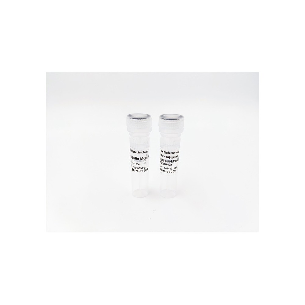

| ELK.No | ES13326 |
| Product name | RSLBA rabbit pAb |
| Reactivity | Human; Mouse;Rat |
| Applications | WB |
| Other name | |
| Size | 50μL |
| Unit price ($) | 148 |
| Human gene ID | 387496 |
| Human Swiss-Prot | Q6T310 |
| Source | Rabbit |
| Isotype | IgG |
| Target | RSLBA |
| Fields | |
| Gene name | RASL11A |
| Protein name | RSLBA |
| Human gene link | View Human Gene |
| Human Swiss link | View Human Swiss-Prot |
| Mouse gene ID | 68895 |
| Mouse gene link | View Mouse Gene |
| Mouse Swiss-Prot | Q6IMB1 |
| Mouse Swiss link | View Mouse Swiss-Prot |
| Rat gene ID | 304268 |
| Rat gene link | View Rat Gene |
| Rat Swiss-Prot | Q6IMA3 |
| Rat Swiss link | View Rat Swiss-Prot |
| Immunogen | Synthesized peptide derived from human RSLBA AA range: 68-118 |
| Specificity | This antibody detects endogenous levels of RSLBA at Human/Mouse/Rat |
| Formulation | Liquid in PBS containing 50% glycerol, 0.5% BSA and 0.02% sodium azide. |
| Clonality | Polyclonal |
| Dilution | WB 1:500-2000 |
| Purification | The antibody was affinity-purified from rabbit serum by affinity-chromatography using specific immunogen. |
| Concentration | 1 mg/ml |
| Storage stability | -20°C/1 year |
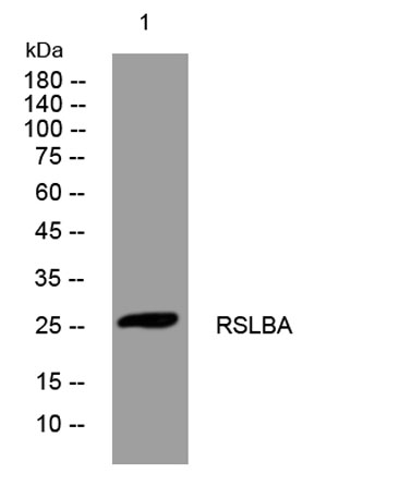
| Molecular Weight (Da) | 27kD |
| Observed band (KD) | |
| Background | RASL11A is a member of the small GTPase protein family with a high degree of similarity to RAS (see HRAS, MIM 190020) proteins.[supplied by OMIM, Nov 2008], |
| Function | developmental stage:Down-regulated during development of THP-1 monocytes into macrophages.,induction:Down-regulated by TGFB1.,similarity:Belongs to the small GTPase superfamily. Ras family.,tissue specificity:Widely expressed. Down-regulated in prostate tumors compared to normal prostate tissue. High levels found in colon tumor and normal colon tissue followed by small intestine, liver, jejunum, ileum, bladder and aorta. Lowest levels observed in endothelial cells., |
| Subcellular location | Nucleus, nucleolus . Associates with rDNA transcription unit throughout the cell cycle. . |
| Expression | Widely expressed. Down-regulated in prostate tumors compared to normal prostate tissue. High levels found in colon tumor and normal colon tissue followed by small intestine, liver, jejunum, ileum, bladder and aorta. Lowest levels observed in endothelial cells. |

Use collapsible tabs for more detailed information that will help customers make a purchasing decision.
Ex: Shipping and return policies, size guides, and other common questions.
