 Français
 English

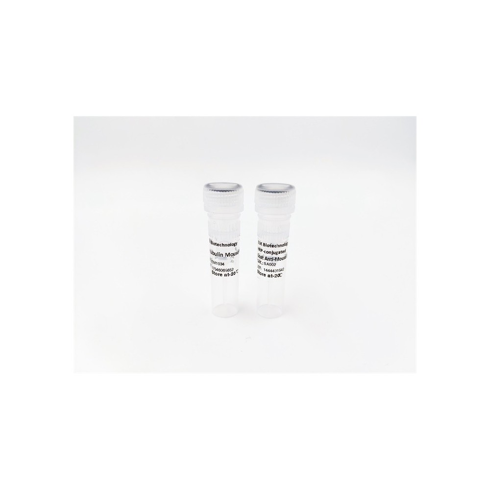

| ELK.No | ES14919 |
| Product name | MOES rabbit pAb |
| Reactivity | Human; Mouse;Rat |
| Applications | WB |
| Other name | |
| Size | 100μL |
| Unit price ($) | 248 |
| Human gene ID | 4478 |
| Human Swiss-Prot | P26038 |
| Source | Rabbit |
| Isotype | IgG |
| Target | MOES |
| Fields | >>Tight junction;>>Leukocyte transendothelial migration;>>Regulation of actin cytoskeleton;>>Measles;>>Proteoglycans in cancer |
| Gene name | MSN |
| Protein name | MOES |
| Human gene link | View Human Gene |
| Human Swiss link | View Human Swiss-Prot |
| Mouse gene ID | 17698 |
| Mouse gene link | View Mouse Gene |
| Mouse Swiss-Prot | P26041 |
| Mouse Swiss link | View Mouse Swiss-Prot |
| Rat gene ID | 81521 |
| Rat gene link | View Rat Gene |
| Rat Swiss-Prot | O35763 |
| Rat Swiss link | View Rat Swiss-Prot |
| Immunogen | Synthesized peptide derived from human MOES AA range: 357-407 |
| Specificity | This antibody detects endogenous levels of MOES at Human/Mouse/Rat |
| Formulation | Liquid in PBS containing 50% glycerol, 0.5% BSA and 0.02% sodium azide. |
| Clonality | Polyclonal |
| Dilution | WB 1:500-2000 |
| Purification | The antibody was affinity-purified from rabbit serum by affinity-chromatography using specific immunogen. |
| Concentration | 1 mg/ml |
| Storage stability | -20°C/1 year |
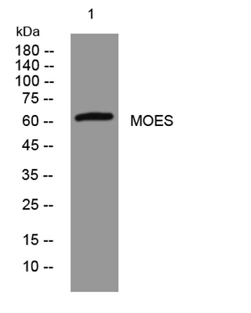
| Molecular Weight (Da) | 63kD |
| Observed band (KD) | |
| Background | Moesin (for membrane-organizing extension spike protein) is a member of the ERM family which includes ezrin and radixin. ERM proteins appear to function as cross-linkers between plasma membranes and actin-based cytoskeletons. Moesin is localized to filopodia and other membranous protrusions that are important for cell-cell recognition and signaling and for cell movement. [provided by RefSeq, Jul 2008], |
| Function | function:Probably involved in connections of major cytoskeletal structures to the plasma membrane.,PTM:Phosphorylation on Thr-558 is crucial for the formation of microvilli-like structures.,similarity:Contains 1 FERM domain.,subcellular location:Phosphorylated form is enriched in microvilli-like structures at apical membrane.,subunit:In resting T-cells, part of a PAG1-SLC9A3R1-MSN complex which is disrupted upon TCR activation (By similarity). Binds SLC9A3R1.,tissue specificity:In all tissues and cultured cells studied., |
| Subcellular location | Cell membrane ; Peripheral membrane protein ; Cytoplasmic side . Cytoplasm, cytoskeleton . Apical cell membrane ; Peripheral membrane protein ; Cytoplasmic side . Cell projection, microvillus membrane ; Peripheral membrane protein ; Cytoplasmic side . Cell projection, microvillus . Phosphorylated form is enriched in microvilli-like structures at apical membrane. Increased cell membrane localization of both phosphorylated and non-phosphorylated forms seen after thrombin treatment (By similarity). Localizes at the uropods of T lymphoblasts. . |
| Expression | In all tissues and cultured cells studied. |

Use collapsible tabs for more detailed information that will help customers make a purchasing decision.
Ex: Shipping and return policies, size guides, and other common questions.