 Français
 English

| ELK.No | ES15063 |
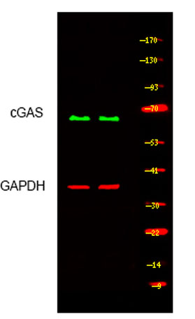
| Product name | cGAS rabbit pAb |
| Reactivity | Human;Rat;Mouse; |
| Applications | WB |
| Other name | |
| Size | 50μL |
| Unit price ($) | 148 |
| Human gene ID | 115004 |
| Human Swiss-Prot | Q8N884 |
| Source | Rabbit |
| Isotype | IgG |
| Target | M21D1 |
| Fields | >>Cytosolic DNA-sensing pathway;>>Shigellosis;>>Human cytomegalovirus infection;>>Herpes simplex virus 1 infection;>>Human immunodeficiency virus 1 infection;>>Coronavirus disease - COVID-19 |
| Gene name | MB21D1 cGAS C6orf150 |
| Protein name | M21D1 |
| Human gene link | View Human Gene |
| Human Swiss link | View Human Swiss-Prot |
| Mouse gene ID | |
| Mouse gene link | |
| Mouse Swiss-Prot | |
| Mouse Swiss link | |
| Rat gene ID | |
| Rat gene link | |
| Rat Swiss-Prot | |
| Rat Swiss link | |
| Immunogen | Synthesized peptide derived from human M21D1 AA range: 35-85 |
| Specificity | This antibody detects endogenous levels of M21D1 at Human |
| Formulation | Liquid in PBS containing 50% glycerol, 0.5% BSA and 0.02% sodium azide. |
| Clonality | Polyclonal |
| Dilution | WB 1:500-2000 |
| Purification | The antibody was affinity-purified from rabbit serum by affinity-chromatography using specific immunogen. |
| Concentration | 1 mg/ml |
| Storage stability | -20°C/1 year |
| Molecular Weight (Da) | 57kD |
| Observed band (KD) | |
| Background | |
| Function | |
| Subcellular location | Nucleus . Chromosome . Cell membrane ; Peripheral membrane protein . Cytoplasm, cytosol . Mainly localizes in the nucleus, and at low level in the cytosol (PubMed:31808743, PubMed:31544964). On chromosomes, enriched on centromeric satellite and LINE DNA repeat elements (PubMed:30811988). Exported from the nucleus to the cytosol in a XPO1/CRM1 via the nuclear export signal in response to DNA stimulation (PubMed:33406424). Outside the nucleus, localizes at the cell membrane as a peripheral membrane protein in resting conditions: association to the cell membrane is mediated via binding to phosphatidylinositol 4,5-bisphosphate (PtdIns(4,5)P2) (PubMed:30827685). Localization at the cell membrane is required to limit the recognition of self-DNA (PubMed:30827685). Following detection of double-st |
| Expression | Expressed in the monocytic cell line THP1. |

Use collapsible tabs for more detailed information that will help customers make a purchasing decision.
Ex: Shipping and return policies, size guides, and other common questions.