 Français
 English

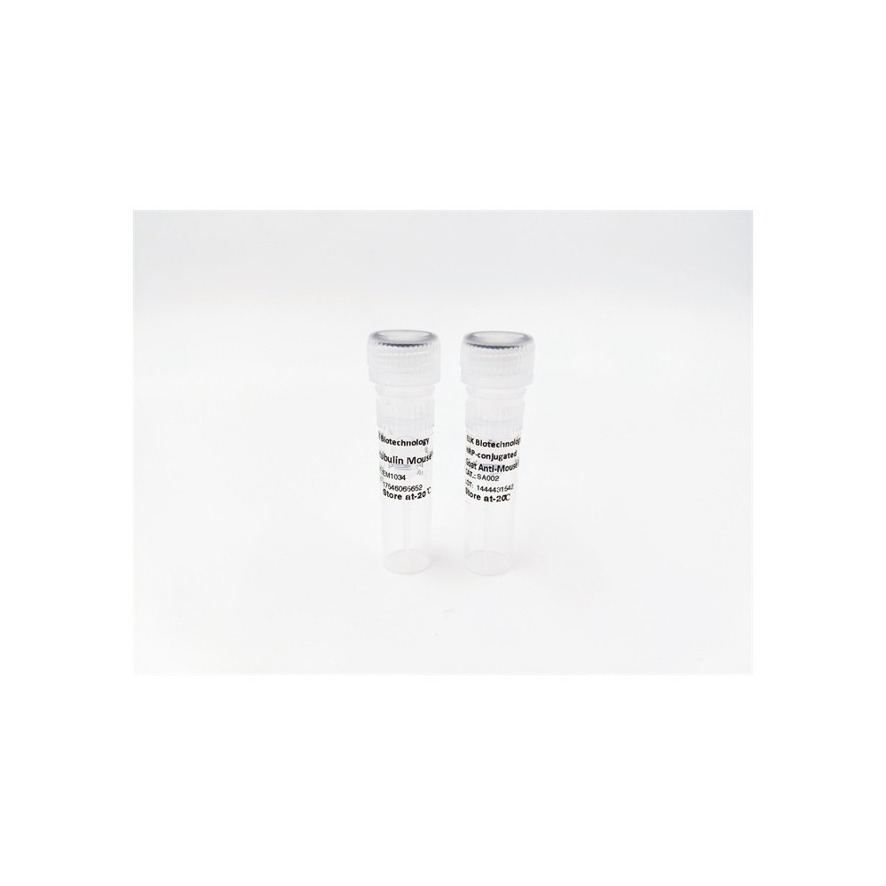

| ELK.No | ES15133 |
| Product name | LOX15 rabbit pAb |
| Reactivity | Human;Rat;Mouse; |
| Applications | WB;ELISA;IHC |
| Other name | |
| Size | 50μL |
| Unit price ($) | 148 |
| Human gene ID | 246 |
| Human Swiss-Prot | P16050 |
| Source | Rabbit |
| Isotype | IgG |
| Target | LOX15 |
| Fields | >>Arachidonic acid metabolism;>>Linoleic acid metabolism;>>Metabolic pathways;>>Ferroptosis;>>Necroptosis;>>Serotonergic synapse |
| Gene name | ALOX15 LOG15 |
| Protein name | LOX15 |
| Human gene link | View Human Gene |
| Human Swiss link | View Human Swiss-Prot |
| Mouse gene ID | |
| Mouse gene link | |
| Mouse Swiss-Prot | |
| Mouse Swiss link | |
| Rat gene ID | |
| Rat gene link | |
| Rat Swiss-Prot | |
| Rat Swiss link | |
| Immunogen | Synthesized peptide derived from human LOX15 AA range: 156-206 |
| Specificity | This antibody detects endogenous levels of LOX15 at Human |
| Formulation | Liquid in PBS containing 50% glycerol, 0.5% BSA and 0.02% sodium azide. |
| Clonality | Polyclonal |
| Dilution | WB 1:500-2000;IHC-p 1:50-300; ELISA 2000-20000 |
| Purification | The antibody was affinity-purified from rabbit serum by affinity-chromatography using specific immunogen. |
| Concentration | 1 mg/ml |
| Storage stability | -20°C/1 year |
| Molecular Weight (Da) | 73kD |
| Observed band (KD) | |
| Background | |
| Function | catalytic activity:Arachidonate + O(2) = (5Z,8Z,11Z,13E)-(15S)-15-hydroperoxyicosa-5,8,11,13-tetraenoate.,cofactor:Binds 1 iron ion per subunit.,function:Converts arachidonic acid to 15S-hydroperoxyeicosatetraenoic acid. Also acts on C-12 of arachidonate as well as on linoleic acid.,pathway:Lipid metabolism; leukotriene D4 biosynthesis.,similarity:Belongs to the lipoxygenase family.,similarity:Contains 1 lipoxygenase domain.,similarity:Contains 1 PLAT domain., |
| Subcellular location | Cytoplasm, cytosol . Cell membrane ; Peripheral membrane protein . Lipid droplet . Predominantly cytosolic; becomes enriched at membranes upon calcium binding (By similarity). Translocates from the cytosol to the plasma membrane when stimulated by IL13/interleukin-13 and in macrophages binding apoptotic cells (By similarity). . |
| Expression | Detected in monocytes and eosinophils (at protein level). Expressed in airway epithelial cells. |



Use collapsible tabs for more detailed information that will help customers make a purchasing decision.
Ex: Shipping and return policies, size guides, and other common questions.