 Français
 English


| ELK.No | ES15338 |
| Product name | KDIS rabbit pAb |
| Reactivity | Human;Rat |
| Applications | WB |
| Other name | |
| Size | 100μL |
| Unit price ($) | 248 |
| Human gene ID | 57498 |
| Human Swiss-Prot | Q9ULH0 |
| Source | Rabbit |
| Isotype | IgG |
| Target | KDIS |
| Fields | >>Neurotrophin signaling pathway |
| Gene name | KIDINS220 ARMS KIAA1250 |
| Protein name | KDIS |
| Human gene link | View Human Gene |
| Human Swiss link | View Human Swiss-Prot |
| Mouse gene ID | |
| Mouse gene link | |
| Mouse Swiss-Prot | |
| Mouse Swiss link | |
| Rat gene ID | 116478 |
| Rat gene link | View Rat Gene |
| Rat Swiss-Prot | Q9EQG6 |
| Rat Swiss link | View Rat Swiss-Prot |
| Immunogen | Synthesized peptide derived from human KDIS AA range: 1023-1073 |
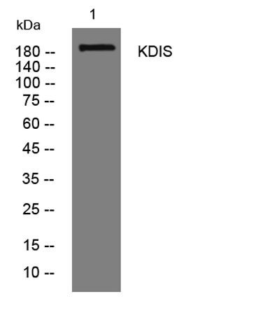
| Specificity | This antibody detects endogenous levels of KDIS at Human/Rat |
| Formulation | Liquid in PBS containing 50% glycerol, 0.5% BSA and 0.02% sodium azide. |
| Clonality | Polyclonal |
| Dilution | WB 1:500-2000 |
| Purification | The antibody was affinity-purified from rabbit serum by affinity-chromatography using specific immunogen. |
| Concentration | 1 mg/ml |
| Storage stability | -20°C/1 year |
| Molecular Weight (Da) | 195kD |
| Observed band (KD) | |
| Background | |
| Function | domain:The transmembrane domain mediates interaction with NTRK1.,function:Promotes a prolonged MAP-kinase signaling by neurotrophins through activation of a Rap1-dependent mechanism. Provides a docking site for the CRKL-C3G complex, resulting in Rap1-dependent sustained ERK activation. May play an important role in regulating postsynaptic signal transduction through the syntrophin-mediated localization of receptor tyrosine kinases such as EPHA4. In cooperation with SNTA1 can enhance EPHA4-induced JAK/STAT activation. May play a role in neurotrophin- and ephrin-mediated neuronal outgrowth and in axon guidance during neural development and in neuronal regeneration (By similarity). Modulates stress-induced apoptosis of melanoma cells via regulation of the MEK/ERK signaling pathway.,PTM:Tyrosine phosphorylated by NTRK1, NTRK2, EPHB2 and EPHA4. Phosphorylation at Ser-918 is induced by phorbol |
| Subcellular location | Membrane ; Multi-pass membrane protein . Late endosome . Localized at late endosome before or after nerve growth factor (NGF) stimulation. |
| Expression | Abundant in developing and adult neural tissues as well as neuroendocrine cells and dendritic cells. Overexpressed in melanoma and melanoma cell lines. |

Use collapsible tabs for more detailed information that will help customers make a purchasing decision.
Ex: Shipping and return policies, size guides, and other common questions.