 Français
 English

IκB-α (phospho-Ser32) rabbit pAb
AO-06-ES15405-100

| ELK.No | ES15405 |
| Product name | IκB-α (phospho-Ser32) rabbit pAb |
| Reactivity | Human;Mouse;Rat |
| Applications | WB;IHC |
| Other name | NF-kappa-B inhibitor alpha (I-kappa-B-alpha) (IkB-alpha) (IkappaBalpha) (Major histocompatibility complex enhancer-binding protein MAD3) |
| Size | 100μL |
| Unit price ($) | 248 |
| Human gene ID | 4792 |
| Human Swiss-Prot | P25963 |
| Source | Rabbit |
| Isotype | IgG |
| Target | IκB-α |
| Fields | >>cAMP signaling pathway;>>Chemokine signaling pathway;>>NF-kappa B signaling pathway;>>Apoptosis;>>Osteoclast differentiation;>>Toll-like receptor signaling pathway;>>NOD-like receptor signaling pathway;>>RIG-I-like receptor signaling pathway;>>Cytosolic DNA-sensing pathway;>>C-type lectin receptor signaling pathway;>>IL-17 signaling pathway;>>Th1 and Th2 cell differentiation;>>Th17 cell differentiation;>>T cell receptor signaling pathway;>>B cell receptor signaling pathway;>>TNF signaling pathway;>>Neurotrophin signaling pathway;>>Adipocytokine signaling pathway;>>Relaxin signaling pathway;>>Insulin resistance;>>Alcoholic liver disease;>>Epithelial cell signaling in Helicobacter pylori infection;>>Pathogenic Escherichia coli infection;>>Shigellosis;>>Salmonella infection;>>Legionellosis;>>Yersinia infection;>>Leishmaniasis;>>Chagas disease;>>Toxoplasmosis;>>Hepatitis C;>>Hepatitis B;>>Measles;>>Human cytomegalovirus infection;>>Influenza A;>>Human T-cell leukemia virus 1 infection;>> |
| Gene name | NFKBIA IKBA MAD3 NFKBI |
| Protein name | NF-kappa-B inhibitor alpha |
| Human gene link | View Human Gene |
| Human Swiss link | View Human Swiss-Prot |
| Mouse gene ID | 18035 |
| Mouse gene link | View Mouse Gene |
| Mouse Swiss-Prot | Q9Z1E3 |
| Mouse Swiss link | View Mouse Swiss-Prot |
| Rat gene ID | 25493 |
| Rat gene link | View Rat Gene |
| Rat Swiss-Prot | Q63746 |
| Rat Swiss link | View Rat Swiss-Prot |
| Immunogen | Synthesized phosho peptide around human IκBα (Ser32) |
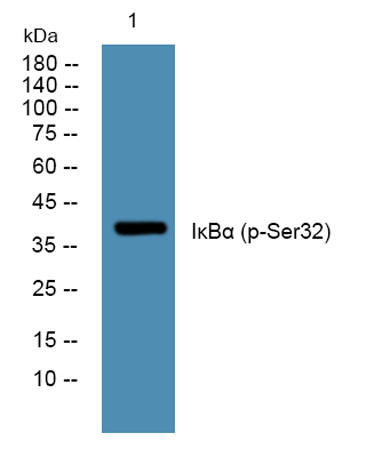
| Specificity | This antibody detects endogenous levels of Human Mouse Rat IκBα (phospho-Ser32) |
| Formulation | Liquid in PBS containing 50% glycerol, 0.5% BSA and 0.02% sodium azide. |
| Clonality | Polyclonal |
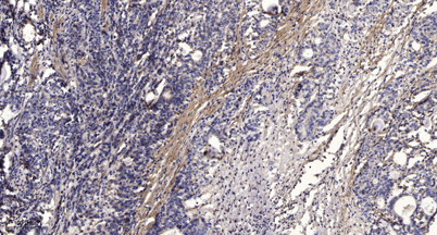
| Dilution | WB 1:500-2000;IHC-p 1:50-300 |
| Purification | The antibody was affinity-purified from rabbit serum by affinity-chromatography using specific immunogen. |
| Concentration | 1 mg/ml |
| Storage stability | -20°C/1 year |
| Molecular Weight (Da) | |
| Observed band (KD) | about 40kd |
| Background | This gene encodes a member of the NF-kappa-B inhibitor family, which contain multiple ankrin repeat domains. The encoded protein interacts with REL dimers to inhibit NF-kappa-B/REL complexes which are involved in inflammatory responses. The encoded protein moves between the cytoplasm and the nucleus via a nuclear localization signal and CRM1-mediated nuclear export. Mutations in this gene have been found in ectodermal dysplasia anhidrotic with T-cell immunodeficiency autosomal dominant disease. [provided by RefSeq, Aug 2011], |
| Function | disease:Defects in NFKBIA are the cause of ectodermal dysplasia anhidrotic with T-cell immunodeficiency autosomal dominant (ADEDAID) [MIM:612132]. Ectodermal dysplasia defines a heterogeneous group of disorders due to abnormal development of two or more ectodermal structures. ADEDAID is an ectodermal dysplasia associated with decreased production of pro-inflammatory cytokines and certain interferons, rendering patients susceptible to infection.,function:Inhibits the activity of dimeric NF-kappa-B/REL complexes by trapping REL dimers in the cytoplasm through masking of their nuclear localization signals. On cellular stimulation by immune and proinflammatory responses, becomes phosphorylated promoting ubiquitination and degradation, enabling the dimeric RELA to tranlocate to the nucleus and activate transcription.,induction:Induced in adherent monocytes.,online information:NFKBIA mutation |
| Subcellular location | Cytoplasm. Nucleus. Shuttles between the nucleus and the cytoplasm by a nuclear localization signal (NLS) and a CRM1-dependent nuclear export. . |
| Expression | Brain,Kidney,Lymph node,Monocyte, |

Use collapsible tabs for more detailed information that will help customers make a purchasing decision.
Ex: Shipping and return policies, size guides, and other common questions.