 Français
 English

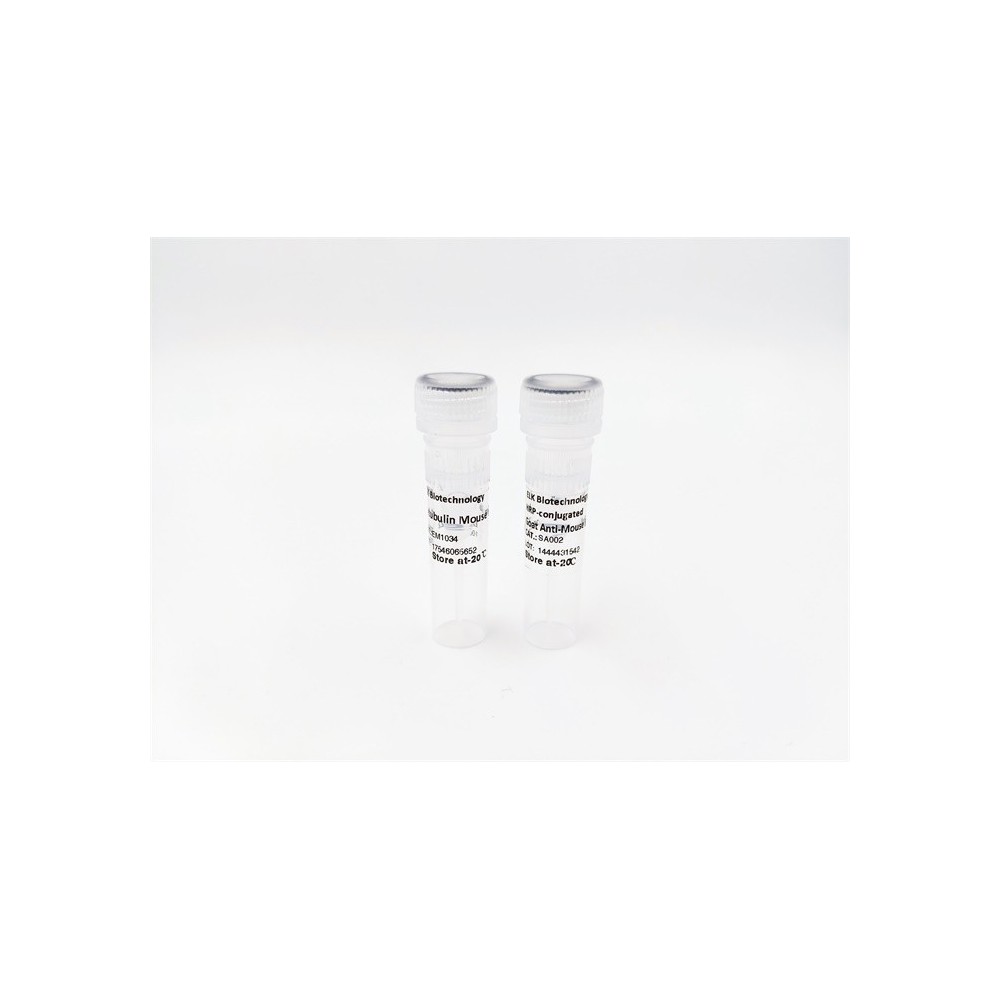

| ELK.No | ES15438 |
| Product name | IPYR rabbit pAb |
| Reactivity | Human; Mouse |
| Applications | WB |
| Other name | |
| Size | 100μL |
| Unit price ($) | 248 |
| Human gene ID | 5464 |
| Human Swiss-Prot | Q15181 |
| Source | Rabbit |
| Isotype | IgG |
| Target | IPYR |
| Fields | >>Oxidative phosphorylation |
| Gene name | PPA1 IOPPP PP |
| Protein name | IPYR |
| Human gene link | View Human Gene |
| Human Swiss link | View Human Swiss-Prot |
| Mouse gene ID | 67895 |
| Mouse gene link | View Mouse Gene |
| Mouse Swiss-Prot | Q9D819 |
| Mouse Swiss link | View Mouse Swiss-Prot |
| Rat gene ID | |
| Rat gene link | |
| Rat Swiss-Prot | |
| Rat Swiss link | |
| Immunogen | Synthesized peptide derived from human IPYR AA range: 162-212 |
| Specificity | This antibody detects endogenous levels of IPYR at Human/Mouse |
| Formulation | Liquid in PBS containing 50% glycerol, 0.5% BSA and 0.02% sodium azide. |
| Clonality | Polyclonal |
| Dilution | WB 1:500-2000 |
| Purification | The antibody was affinity-purified from rabbit serum by affinity-chromatography using specific immunogen. |
| Concentration | 1 mg/ml |
| Storage stability | -20°C/1 year |
| Molecular Weight (Da) | 32kD |
| Observed band (KD) | |
| Background | The protein encoded by this gene is a member of the inorganic pyrophosphatase (PPase) family. PPases catalyze the hydrolysis of pyrophosphate to inorganic phosphate, which is important for the phosphate metabolism of cells. Studies of a similar protein in bovine suggested a cytoplasmic localization of this enzyme. [provided by RefSeq, Jul 2008], |
| Function | catalytic activity:Diphosphate + H(2)O = 2 phosphate.,cofactor:Binds 4 magnesium ions per subunit. Other metal ions can support activity, but at a lower rate. Two magnesium ions are required for the activation of the enzyme and are present before substrate binds, two additional magnesium ions form complexes with substrate and product.,similarity:Belongs to the PPase family.,subunit:Homodimer.,tissue specificity:Expressed ubiquitously., |
| Subcellular location | Cytoplasm . |
| Expression | Expressed ubiquitously. |


Use collapsible tabs for more detailed information that will help customers make a purchasing decision.
Ex: Shipping and return policies, size guides, and other common questions.