 Français
 English

| ELK.No | ES1648 |
| Product name | AMPKα1 rabbit pAb |
| Reactivity | Human;Mouse;Rat |
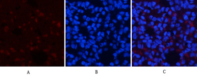
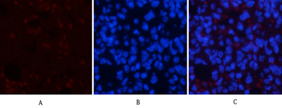
| Applications | WB;IHC;IF;ELISA |
| Other name | PRKAA1; AMPK1; 5'-AMP-activated protein kinase catalytic subunit alpha-1; AMPK subunit alpha-1; Acetyl-CoA carboxylase kinase; ACACA kinase; Hydroxymethylglutaryl-CoA reductase kinase; HMGCR kinase; Tau-protein kinase PRKAA1 |
| Size | 50μL |
| Unit price ($) | 148 |
| Human gene ID | 5562 |
| Human Swiss-Prot | Q13131 |
| Source | Rabbit |
| Isotype | IgG |
| Target | AMPKα1 |
| Fields | >>FoxO signaling pathway;>>Autophagy - animal;>>mTOR signaling pathway;>>PI3K-Akt signaling pathway;>>AMPK signaling pathway;>>Longevity regulating pathway;>>Longevity regulating pathway - multiple species;>>Apelin signaling pathway;>>Tight junction;>>Circadian rhythm;>>Thermogenesis;>>Insulin signaling pathway;>>Adipocytokine signaling pathway;>>Oxytocin signaling pathway;>>Glucagon signaling pathway;>>Insulin resistance;>>Non-alcoholic fatty liver disease;>>Alcoholic liver disease;>>Hypertrophic cardiomyopathy;>>Fluid shear stress and atherosclerosis |
| Gene name | PRKAA1 |
| Protein name | 5'-AMP-activated protein kinase catalytic subunit alpha-1 |
| Human gene link | View Human Gene |
| Human Swiss link | View Human Swiss-Prot |
| Mouse gene ID | 105787 |
| Mouse gene link | View Mouse Gene |
| Mouse Swiss-Prot | Q5EG47 |
| Mouse Swiss link | View Mouse Swiss-Prot |
| Rat gene ID | 65248 |
| Rat gene link | View Rat Gene |
| Rat Swiss-Prot | P54645 |
| Rat Swiss link | View Rat Swiss-Prot |
| Immunogen | The antiserum was produced against synthesized peptide derived from human AMPK1. AA range:451-500 |
| Specificity | AMPKα1 Polyclonal Antibody detects endogenous levels of AMPKα1 protein. |
| Formulation | Liquid in PBS containing 50% glycerol, 0.5% BSA and 0.02% sodium azide. |
| Clonality | Polyclonal |
| Dilution | Western Blot: 1/500 - 1/2000. Immunohistochemistry: 1/100 - 1/300. Immunofluorescence: 1/200 - 1/1000. ELISA: 1/10000. Not yet tested in other applications. |
| Purification | The antibody was affinity-purified from rabbit antiserum by affinity-chromatography using epitope-specific immunogen. |
| Concentration | 1 mg/ml |
| Storage stability | -20°C/1 year |
| Molecular Weight (Da) | |
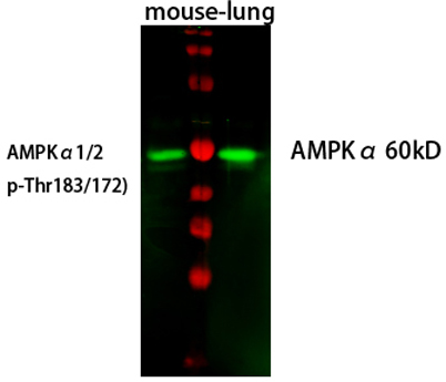
| Observed band (KD) | 65kD |
| Background | The protein encoded by this gene belongs to the ser/thr protein kinase family. It is the catalytic subunit of the 5'-prime-AMP-activated protein kinase (AMPK). AMPK is a cellular energy sensor conserved in all eukaryotic cells. The kinase activity of AMPK is activated by the stimuli that increase the cellular AMP/ATP ratio. AMPK regulates the activities of a number of key metabolic enzymes through phosphorylation. It protects cells from stresses that cause ATP depletion by switching off ATP-consuming biosynthetic pathways. Alternatively spliced transcript variants encoding distinct isoforms have been observed. [provided by RefSeq, Jul 2008], |
| Function | catalytic activity:ATP + a protein = ADP + a phosphoprotein.,cofactor:Magnesium.,enzyme regulation:Binding of AMP results in allosteric activation, inducing phosphorylation on Thr-174 by STK11 in complex with STE20-related adapter-alpha (STRAD alpha) pseudo kinase and CAB39. Also activated by phosphorylation by CAMKK2 triggered by a rise in intracellular calcium ions, without detectable changes in the AMP/ATP ratio.,function:Responsible for the regulation of fatty acid synthesis by phosphorylation of acetyl-CoA carboxylase. It also regulates cholesterol synthesis via phosphorylation and inactivation of hormone-sensitive lipase and hydroxymethylglutaryl-CoA reductase. Appears to act as a metabolic stress-sensing protein kinase switching off biosynthetic pathways when cellular ATP levels are depleted and when 5'-AMP rises in response to fuel limitation and/or hypoxia. This is a catalytic s |
| Subcellular location | Cytoplasm . Nucleus . In response to stress, recruited by p53/TP53 to specific promoters. . |
| Expression | Brain,Intestine,Liver,Mammary gland,Platelet,Testis |

Use collapsible tabs for more detailed information that will help customers make a purchasing decision.
Ex: Shipping and return policies, size guides, and other common questions.