 Français
 English

| ELK.No | ES1674 |
| Product name | APC rabbit pAb |
| Reactivity | Human;Mouse;Rat |
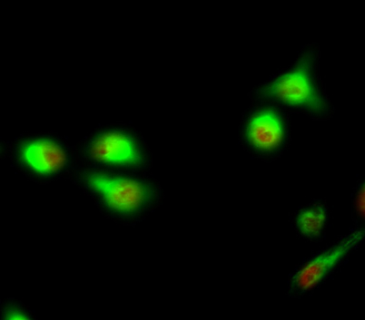
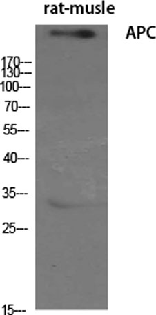
| Applications | WB;IHC;IF;ELISA |
| Other name | APC; DP2.5; Adenomatous polyposis coli protein; Protein APC; Deleted in polyposis 2.5 |
| Size | 100μL |
| Unit price ($) | 248 |
| Human gene ID | 324 |
| Human Swiss-Prot | P25054 |
| Source | Rabbit |
| Isotype | IgG |
| Target | APC |
| Fields | >>Wnt signaling pathway;>>Hippo signaling pathway;>>Signaling pathways regulating pluripotency of stem cells;>>Regulation of actin cytoskeleton;>>Cushing syndrome;>>Alzheimer disease;>>Pathways of neurodegeneration - multiple diseases;>>Human papillomavirus infection;>>Pathways in cancer;>>MicroRNAs in cancer;>>Colorectal cancer;>>Endometrial cancer;>>Basal cell carcinoma;>>Breast cancer;>>Hepatocellular carcinoma;>>Gastric cancer |
| Gene name | APC |
| Protein name | Adenomatous polyposis coli protein |
| Human gene link | View Human Gene |
| Human Swiss link | View Human Swiss-Prot |
| Mouse gene ID | |
| Mouse gene link | |
| Mouse Swiss-Prot | Q61315 |
| Mouse Swiss link | View Mouse Swiss-Prot |
| Rat gene ID | 24205 |
| Rat gene link | View Rat Gene |
| Rat Swiss-Prot | P70478 |
| Rat Swiss link | View Rat Swiss-Prot |
| Immunogen | The antiserum was produced against synthesized peptide derived from human APC. AA range:2794-2843 |
| Specificity | APC Polyclonal Antibody detects endogenous levels of APC protein. |
| Formulation | Liquid in PBS containing 50% glycerol, 0.5% BSA and 0.02% sodium azide. |
| Clonality | Polyclonal |
| Dilution | Western Blot: 1/500 - 1/2000. Immunohistochemistry: 1/100 - 1/300. Immunofluorescence: 1/200 - 1/1000. ELISA: 1/40000. Not yet tested in other applications. |
| Purification | The antibody was affinity-purified from rabbit antiserum by affinity-chromatography using epitope-specific immunogen. |
| Concentration | 1 mg/ml |
| Storage stability | -20°C/1 year |
| Molecular Weight (Da) | |
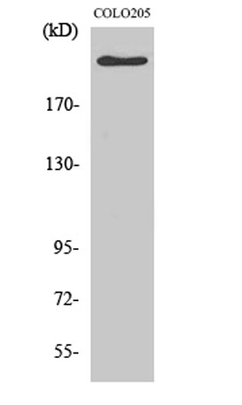
| Observed band (KD) | 310kD |
| Background | This gene encodes a tumor suppressor protein that acts as an antagonist of the Wnt signaling pathway. It is also involved in other processes including cell migration and adhesion, transcriptional activation, and apoptosis. Defects in this gene cause familial adenomatous polyposis (FAP), an autosomal dominant pre-malignant disease that usually progresses to malignancy. Disease-associated mutations tend to be clustered in a small region designated the mutation cluster region (MCR) and result in a truncated protein product. [provided by RefSeq, Jul 2008], |
| Function | disease:APC mutations have led to some interesting observations. (1) the great majority of the mutations found to date would result in truncation of the APC product. (2) almost all the mutations have occurred within the first half of the coding sequence, and somatic mutations in colorectal tumors are further clustered in a particular region, called MCR (mutation cluster region). (3) most identified point mutations in the APC gene are transitions from cytosine to other nucleotides. (4) the location of germline mutations tends to correlate with the number of colorectal polyps in FAP patients. Inactivation of both alleles of the APC gene seems to be required as an early event to develop most adenomas and carcinomas in the colon and rectum as well as some of those in the stomach.,disease:Defects in APC are a cause of familial adenomatous polyposis (FAP) [MIM:175100]; which includes also Gard |
| Subcellular location | Cell junction, adherens junction . Cytoplasm, cytoskeleton . Cell projection, lamellipodium . Cell projection, ruffle membrane . Cytoplasm . Cell membrane . Associated with the microtubule network at the growing distal tip of microtubules (PubMed:19632184). Accumulates in the lamellipodium and ruffle membrane in response to hepatocyte growth factor (HGF) treatment (PubMed:19151759). The MEMO1-RHOA-DIAPH1 signaling pathway controls localization of the phosphorylated form to the cell membrane (PubMed:20937854). . |
| Expression | Expressed in a variety of tissues: brain, small intestine, colon, thymus, skeletal muscle, heart, prostate, lung, spleen, ovary, testis kidney, placenta, blood and liver (PubMed:21643010, PubMed:27217144). Isoform 1A: Very strongly expressed in brain but has relatively low expression levels in other tissues (PubMed:19527921, PubMed:21643010, PubMed:27217144). Isoform 1B: Predominant form in all tissues except for brain, including gastric mucosa and blood (PubMed:19527921, PubMed:21643010, PubMed:27217144). |

Use collapsible tabs for more detailed information that will help customers make a purchasing decision.
Ex: Shipping and return policies, size guides, and other common questions.