 Français
 English

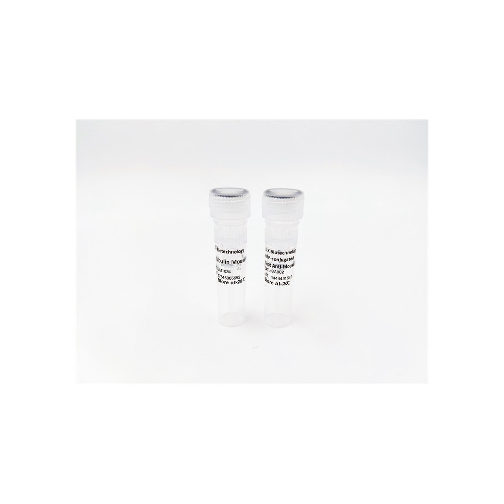

| ELK.No | ES16886 |
| Product name | DTBP1 rabbit pAb |
| Reactivity | Human; Mouse;Rat |
| Applications | WB |
| Other name | |
| Size | 100μL |
| Unit price ($) | 248 |
| Human gene ID | 84062 |
| Human Swiss-Prot | Q96EV8 |
| Source | Rabbit |
| Isotype | IgG |
| Target | DTBP1 |
| Fields | |
| Gene name | DTNBP1 My031 |
| Protein name | DTBP1 |
| Human gene link | View Human Gene |
| Human Swiss link | View Human Swiss-Prot |
| Mouse gene ID | 94245 |
| Mouse gene link | View Mouse Gene |
| Mouse Swiss-Prot | Q91WZ8 |
| Mouse Swiss link | View Mouse Swiss-Prot |
| Rat gene ID | 641528 |
| Rat gene link | View Rat Gene |
| Rat Swiss-Prot | Q5M834 |
| Rat Swiss link | View Rat Swiss-Prot |
| Immunogen | Synthesized peptide derived from human DTBP1 AA range: 56-106 |
| Specificity | This antibody detects endogenous levels of DTBP1 at Human/Mouse/Rat |
| Formulation | Liquid in PBS containing 50% glycerol, 0.5% BSA and 0.02% sodium azide. |
| Clonality | Polyclonal |
| Dilution | WB 1:500-2000 |
| Purification | The antibody was affinity-purified from rabbit serum by affinity-chromatography using specific immunogen. |
| Concentration | 1 mg/ml |
| Storage stability | -20°C/1 year |
| Molecular Weight (Da) | 39kD |
| Observed band (KD) | |
| Background | This gene encodes a protein that may play a role in organelle biogenesis associated with melanosomes, platelet dense granules, and lysosomes. A similar protein in mouse is a component of a protein complex termed biogenesis of lysosome-related organelles complex 1 (BLOC-1), and binds to alpha- and beta-dystrobrevins, which are components of the dystrophin-associated protein complex (DPC). Mutations in this gene are associated with Hermansky-Pudlak syndrome type 7. This gene may also be associated with schizophrenia. Multiple transcript variants encoding distinct isoforms have been identified for this gene. [provided by RefSeq, Jul 2008], |
| Function | caution:The sequence shown here is derived from an Ensembl automatic analysis pipeline and should be considered as preliminary data.,disease:Defects in DTNBP1 are the cause of Hermansky-Pudlak syndrome type 7 (HPS7) [MIM:203300]. Hermansky-Pudlak syndrome (HPS) is a genetically heterogeneous, rare, autosomal recessive disorder characterized by oculocutaneous albinism, bleeding due to platelet storage pool deficiency, and lysosomal storage defects. This syndrome results from defects of diverse cytoplasmic organelles including melanosomes, platelet dense granules and lysosomes. Ceroid storage in the lungs is associated with pulmonary fibrosis, a common cause of premature death in individuals with HPS.,function:Plays a role in the biogenesis of lysosome-related organelles such as platelet dense granule and melanosomes.,similarity:Belongs to the dysbindin family.,subcellular location:Associa |
| Subcellular location | [Isoform 1]: Cytoplasm . Cytoplasmic vesicle membrane ; Peripheral membrane protein ; Cytoplasmic side . Endosome membrane ; Peripheral membrane protein ; Cytoplasmic side . Melanosome membrane ; Peripheral membrane protein ; Cytoplasmic side . Cell junction, synapse, postsynaptic density . Endoplasmic reticulum . Nucleus . Mainly cytoplasmic but shuttles between the cytoplasm and nucleus. Exported out of the nucleus via its NES in a XPO1-dependent manner. Nuclear localization is required for regulation of the expression of genes such as SYN1. Detected in neuron cell bodies, axons and dendrites. Mainly located to the postsynaptic density. Detected at tubulovesicular elements in the vicinity of the Golgi apparatus and of melanosomes. Occasionally detected at the membrane of pigmented melano |
| Expression | Detected in brain, in neurons and in neuropil. Isoform 1 is expressed in the cerebral cortex, and hippocampal frontal (HF). Specific expression in the posterior half of the superior temporal gyrus (pSTG). Higher expression of isoform 2 and 3 in the HF than in the pSTG while isoform 1 shows no difference in expression in these areas. In the HF, detected in dentate gyrus (DG) and in pyramidal cells of hippocampus CA2 and CA3 (at protein level). Expressed in all principal neuronal populations of the HF, namely pyramidal neurons in the subiculum and CA1-3, granule cells in the dense cell layer of the DG (DGg), and polymorph cells in the hilus of the DG (DGh). Maximal levels in CA2, CA3, and DGh. Isoform 2 not expressed in the cerebral cortex. |


Use collapsible tabs for more detailed information that will help customers make a purchasing decision.
Ex: Shipping and return policies, size guides, and other common questions.