 Français
 English
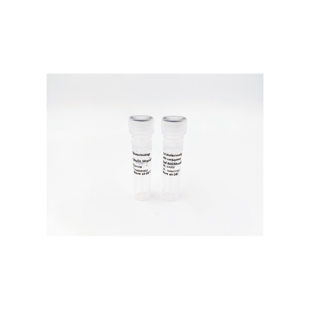

| ELK.No | ES17381 |
| Product name | CLCN3 rabbit pAb |
| Reactivity | Human; Mouse;Rat |
| Applications | WB |
| Other name | |
| Size | 50μL |
| Unit price ($) | 148 |
| Human gene ID | 1182 |
| Human Swiss-Prot | P51790 |
| Source | Rabbit |
| Isotype | IgG |
| Target | CLCN3 |
| Fields | >>Neutrophil extracellular trap formation |
| Gene name | CLCN3 |
| Protein name | CLCN3 |
| Human gene link | View Human Gene |
| Human Swiss link | View Human Swiss-Prot |
| Mouse gene ID | 12725 |
| Mouse gene link | View Mouse Gene |
| Mouse Swiss-Prot | P51791 |
| Mouse Swiss link | View Mouse Swiss-Prot |
| Rat gene ID | 84360 |
| Rat gene link | View Rat Gene |
| Rat Swiss-Prot | P51792 |
| Rat Swiss link | View Rat Swiss-Prot |
| Immunogen | Synthesized peptide derived from human CLCN3 AA range: 597-647 |
| Specificity | This antibody detects endogenous levels of CLCN3 at Human/Mouse/Rat |
| Formulation | Liquid in PBS containing 50% glycerol, 0.5% BSA and 0.02% sodium azide. |
| Clonality | Polyclonal |
| Dilution | WB 1:500-2000 |
| Purification | The antibody was affinity-purified from rabbit serum by affinity-chromatography using specific immunogen. |
| Concentration | 1 mg/ml |
| Storage stability | -20°C/1 year |
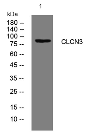
| Molecular Weight (Da) | 90kD |
| Observed band (KD) | |
| Background | This gene encodes a member of the voltage-gated chloride channel (ClC) family. The encoded protein is present in all cell types and localized in plasma membranes and in intracellular vesicles. It is a multi-pass membrane protein which contains a ClC domain and two additional C-terminal CBS (cystathionine beta-synthase) domains. The ClC domain catalyzes the selective flow of Cl- ions across cell membranes, and the CBS domain may have a regulatory function. This protein plays a role in both acidification and transmitter loading of GABAergic synaptic vesicles, and in smooth muscle cell activation and neointima formation. This protein is required for lysophosphatidic acid (LPA)-activated Cl- current activity and fibroblast-to-myofibroblast differentiation. The protein activity is regulated by Ca(2+)/calmodulin-dependent protein kinase II (CaMKII) in glioma cells. Multiple alternatively spliced transcript variants encoding different isoforms have been identified. [provided by RefSeq, Aug 2011], |
| Function | domain:Isoform 2 contains a C-terminal PDZ-binding motif mediating the interaction with GOPC.,function:Mediates the exchange of chloride ions against protons. Functions as antiporter and contributes to the acidification of the endosome and synaptic vesicle lumen, and may thereby affect vesicle trafficking and exocytosis. May play an important role in neuronal cell function through regulation of membrane excitability by protein kinase C. It could help neuronal cells to establish short-term memory.,miscellaneous:The CLC channel family contains both chloride channels and proton-coupled anion transporters that exchange chloride or another anion for protons. The presence of conserved gating glutamate residues is typical for family members that function as antiporters.,PTM:N-glycosylated.,similarity:Belongs to the chloride channel (TC 2.A.49) family.,similarity:Contains 2 CBS domains.,subcellu |
| Subcellular location | [Isoform 1]: Early endosome membrane ; Multi-pass membrane protein . Late endosome membrane ; Multi-pass membrane protein . Lysosome membrane ; Multi-pass membrane protein . Cell membrane ; Multi-pass membrane protein . Isoform 1 is localized mainly in late endosomes. .; [Isoform 2]: Golgi apparatus membrane ; Multi-pass membrane protein . Cell projection, ruffle membrane ; Multi-pass membrane protein . Isoform 2 is mainly enriched in the Golgi (PubMed:12471024). Colocalizes with SLC9A3R1/EBP50 in membrane ruffles (PubMed:11967229). . |
| Expression | Expressed primarily in tissues derived from neuroectoderm. Within the brain, its expression is particularly evident in the hippocampus, olfactory cortex, and olfactory bulb. Highly expressed in aortic and coronary vascular smooth muscle cells, and aortic endothelial cells. Also expressed in tracheal and alveolar epithelial cells, and intima and media of the pulmonary vessels. Expressed in bronchus and colon (at protein level). |

Use collapsible tabs for more detailed information that will help customers make a purchasing decision.
Ex: Shipping and return policies, size guides, and other common questions.