 Français
 English

| ELK.No | ES17518 |
| Product name | CETN1 rabbit pAb |
| Reactivity | Human; Mouse |
| Applications | WB |
| Other name | |
| Size | 50μL |
| Unit price ($) | 148 |
| Human gene ID | 1068 |
| Human Swiss-Prot | Q12798 |
| Source | Rabbit |
| Isotype | IgG |
| Target | CETN1 |
| Fields | |
| Gene name | CETN1 CEN1 CETN |
| Protein name | CETN1 |
| Human gene link | View Human Gene |
| Human Swiss link | View Human Swiss-Prot |
| Mouse gene ID | 26369 |
| Mouse gene link | View Mouse Gene |
| Mouse Swiss-Prot | P41209 |
| Mouse Swiss link | View Mouse Swiss-Prot |
| Rat gene ID | |
| Rat gene link | |
| Rat Swiss-Prot | |
| Rat Swiss link | |
| Immunogen | Synthesized peptide derived from human CETN1 AA range: 96-146 |
| Specificity | This antibody detects endogenous levels of CETN1 at Human/Mouse |
| Formulation | Liquid in PBS containing 50% glycerol, 0.5% BSA and 0.02% sodium azide. |
| Clonality | Polyclonal |
| Dilution | WB 1:500-2000 |
| Purification | The antibody was affinity-purified from rabbit serum by affinity-chromatography using specific immunogen. |
| Concentration | 1 mg/ml |
| Storage stability | -20°C/1 year |
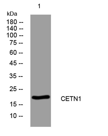
| Molecular Weight (Da) | 19kD |
| Observed band (KD) | |
| Background | The protein encoded by this gene plays important roles in the determination of centrosome position and segregation, and in the process of microtubule severing. This protein is localized to the centrosome of interphase cells, and redistributes to the region of the spindle poles during mitosis, reflecting the dynamic behavior of the centrosome during the cell cycle. [provided by RefSeq, Jan 2015], |
| Function | function:Plays a fundamental role in microtubule-organizing center structure and function.,miscellaneous:Binds two moles of calcium per mole of protein.,similarity:Belongs to the centrin family.,similarity:Contains 4 EF-hand domains.,subcellular location:Centrosome of interphase and mitotic cells.,subunit:Monomer., |
| Subcellular location | Cytoplasm, cytoskeleton, microtubule organizing center, centrosome . Cell projection, cilium . Centrosome of interphase and mitotic cells. In the retinal photoreceptor cells, localizes at the connecting cilium, a thin bridge linking the cell body and the light-sensing outer segment (By similarity). . |
| Expression |

Use collapsible tabs for more detailed information that will help customers make a purchasing decision.
Ex: Shipping and return policies, size guides, and other common questions.