 Français
 English
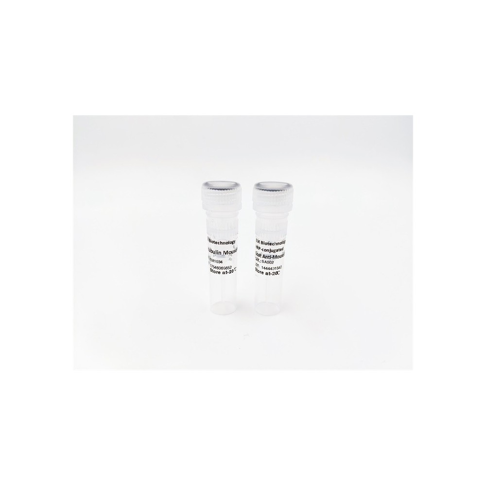

| ELK.No | ES18239 |
| Product name | ASB1 rabbit pAb |
| Reactivity | Human; Mouse |
| Applications | WB |
| Other name | |
| Size | 50μL |
| Unit price ($) | 148 |
| Human gene ID | 51665 |
| Human Swiss-Prot | Q9Y576 |
| Source | Rabbit |
| Isotype | IgG |
| Target | ASB1 |
| Fields | |
| Gene name | ASB1 KIAA1146 |
| Protein name | ASB1 |
| Human gene link | View Human Gene |
| Human Swiss link | View Human Swiss-Prot |
| Mouse gene ID | 65247 |
| Mouse gene link | View Mouse Gene |
| Mouse Swiss-Prot | Q9WV74 |
| Mouse Swiss link | View Mouse Swiss-Prot |
| Rat gene ID | |
| Rat gene link | |
| Rat Swiss-Prot | |
| Rat Swiss link | |
| Immunogen | Synthesized peptide derived from human ASB1 AA range: 76-126 |
| Specificity | This antibody detects endogenous levels of ASB1 at Human/Mouse |
| Formulation | Liquid in PBS containing 50% glycerol, 0.5% BSA and 0.02% sodium azide. |
| Clonality | Polyclonal |
| Dilution | WB 1:500-2000 |
| Purification | The antibody was affinity-purified from rabbit serum by affinity-chromatography using specific immunogen. |
| Concentration | 1 mg/ml |
| Storage stability | -20°C/1 year |
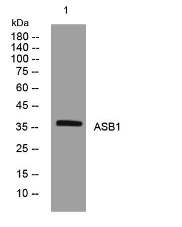
| Molecular Weight (Da) | 37kD |
| Observed band (KD) | |
| Background | The protein encoded by this gene contains an ankyrin repeat sequence and SOCS box domain. The SOCS box serves to couple suppressor of cytokine signalling (SOCS) proteins and their binding partners with the elongin B and C complex, targeting them for ubiquitination and degradation. [provided by RefSeq, Aug 2016], |
| Function | domain:The SOCS box domain mediates the interaction with the Elongin BC complex, an adapter module in different E3 ubiquitin-protein ligase complexes.,function:May play a role in testis development (By similarity). Probable substrate-recognition component of a SCF-like ECS (Elongin-Cullin-SOCS-box protein) E3 ligase complex which mediates the ubiquitination and subsequent proteasomal degradation of target proteins.,pathway:Protein modification; protein ubiquitination.,similarity:Contains 1 SOCS box domain.,similarity:Contains 6 ANK repeats.,subunit:Interacts with CUL5 and RNF7., |
| Subcellular location | intracellular, |
| Expression |

Use collapsible tabs for more detailed information that will help customers make a purchasing decision.
Ex: Shipping and return policies, size guides, and other common questions.