 Français
 English

| ELK.No | ES1881 |
| Product name | CBP 35 rabbit pAb |
| Reactivity | Human;Mouse;Rat |
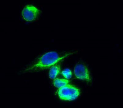
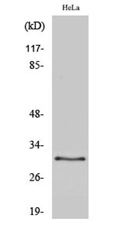
| Applications | WB;IHC;IF;ELISA |
| Other name | LGALS3; MAC2; Galectin-3; Gal-3; 35 kDa lectin; Carbohydrate-binding protein 35; CBP 35; Galactose-specific lectin 3; Galactoside-binding protein; GALBP; IgE-binding protein; L-31; Laminin-binding protein; Lectin L-29; Mac-2 antigen |
| Size | 50μL |
| Unit price ($) | 148 |
| Human gene ID | 3958 |
| Human Swiss-Prot | P17931 |
| Source | Rabbit |
| Isotype | IgG |
| Target | Galectin-3 |
| Fields | |
| Gene name | LGALS3 |
| Protein name | Galectin-3 |
| Human gene link | View Human Gene |
| Human Swiss link | View Human Swiss-Prot |
| Mouse gene ID | |
| Mouse gene link | |
| Mouse Swiss-Prot | P16110 |
| Mouse Swiss link | View Mouse Swiss-Prot |
| Rat gene ID | 83781 |
| Rat gene link | View Rat Gene |
| Rat Swiss-Prot | P08699 |
| Rat Swiss link | View Rat Swiss-Prot |
| Immunogen | The antiserum was produced against synthesized peptide derived from human Galectin 3. AA range:141-190 |
| Specificity | CBP 35 Polyclonal Antibody detects endogenous levels of CBP 35 protein. |
| Formulation | Liquid in PBS containing 50% glycerol, 0.5% BSA and 0.02% sodium azide. |
| Clonality | Polyclonal |
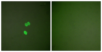
| Dilution | Western Blot: 1/500 - 1/2000. Immunohistochemistry: 1/100 - 1/300. Immunofluorescence: 1/200 - 1/1000. ELISA: 1/20000. Not yet tested in other applications. |
| Purification | The antibody was affinity-purified from rabbit antiserum by affinity-chromatography using epitope-specific immunogen. |
| Concentration | 1 mg/ml |
| Storage stability | -20°C/1 year |
| Molecular Weight (Da) | |
| Observed band (KD) | 33kD |
| Background | This gene encodes a member of the galectin family of carbohydrate binding proteins. Members of this protein family have an affinity for beta-galactosides. The encoded protein is characterized by an N-terminal proline-rich tandem repeat domain and a single C-terminal carbohydrate recognition domain. This protein can self-associate through the N-terminal domain allowing it to bind to multivalent saccharide ligands. This protein localizes to the extracellular matrix, the cytoplasm and the nucleus. This protein plays a role in numerous cellular functions including apoptosis, innate immunity, cell adhesion and T-cell regulation. The protein exhibits antimicrobial activity against bacteria and fungi. Alternate splicing results in multiple transcript variants.[provided by RefSeq, Oct 2014], |
| Function | function:Galactose-specific lectin which binds IgE. May mediate with the alpha-3, beta-1 integrin the stimulation by CSPG4 of endothelial cells migration. Together with DMBT1, required for terminal differentiation of columnar epithelial cells during early embryogenesis.,online information:Galectin-3,similarity:Contains 1 galectin domain.,subcellular location:Cytoplasmic in adenomas and carcinomas. May be secreted by a non-classical secretory pathway and associate with the cell surface.,subunit:Probably forms homo- or heterodimers. Interacts with DMBT1 (By similarity). Forms a complex with the ITGA3, ITGB1 and CSPG4. Interacts with LGALS3BP, LYPD3, CYHR1 and UACA.,tissue specificity:A major expression is found in the colonic epithelium. It is also abundant in the activated macrophages., |
| Subcellular location | Cytoplasm . Nucleus. Secreted . Secreted by a non-classical secretory pathway and associates with the cell surface. Can be secreted; the secretion is dependent on protein unfolding and facilitated by the cargo receptor TMED10; it results in protein translocation from the cytoplasm into the ERGIC (endoplasmic reticulum-Golgi intermediate compartment) followed by vesicle entry and secretion (PubMed:32272059). . |
| Expression | A major expression is found in the colonic epithelium. It is also abundant in the activated macrophages. Expressed in fetal membranes. |

Use collapsible tabs for more detailed information that will help customers make a purchasing decision.
Ex: Shipping and return policies, size guides, and other common questions.